NANOSCOPIUM
Nanoscopium is a unique innovative beamline, which is dedicated to fast scanning multimodal and multi-lengthscale (30 nm à 1µm) X-ray imaging.
It offers simultaneous information in a quantitative manner and in the same experimental conditions, in 2D & 3D (tomography) about the elemental composition, chemical speciation and sample morphology.
The NANOSCOPIUM beamline is open for user proposals
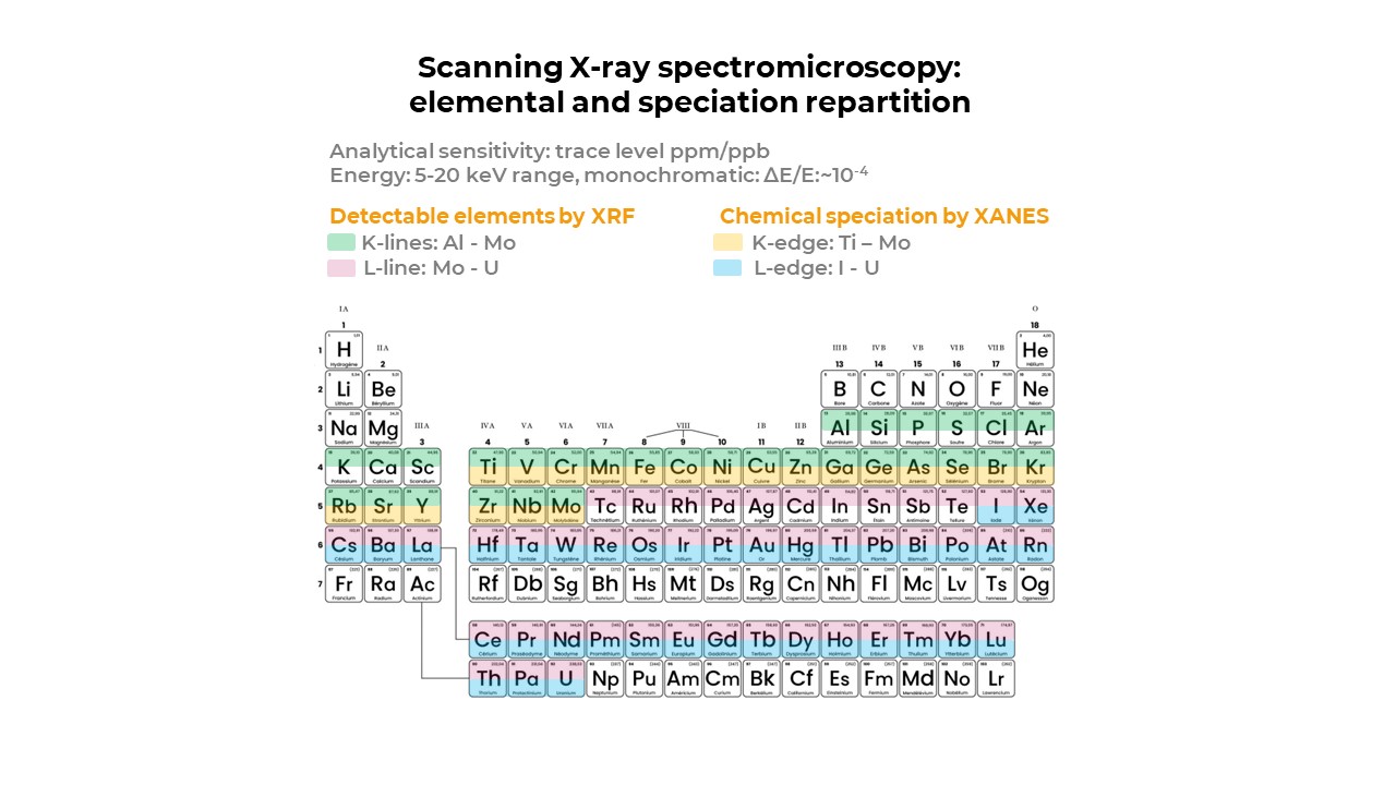
The Nanoscopium hard X-ray (5-20 keV) nanoprobe beamline is dedicated to multi-technique X-ray imaging using fast scanning and high spatial resolution. The beamline develops and offers state-of-the-art X-ray nano-imaging and tomography techniques. Indeed, Nanoscopium offers a large portfolio of complementary imaging and spectroscopy methods, also in association to coherent diffraction imaging techniques. The cutting edge multitechnique possibilities available at the beamline pave the way towards quantitative imaging (morphology, elemental composition and chemical speciation) at hierarchical length-scales (30nm – 1µm).
The actually open User Proposal Call of Synchrotron Soleil can be found at https://www.synchrotron-soleil.fr/en/users.
Scanning multi-lengthscale imaging : the spatial resolution is adaptable in the 50 nanometre - 1 micron range
Complex samples with heterogene composition and tiny features of several millimetres samples can be easily studied by this unique zoom-in capability. The results shown below are published in "Highlights 2018 Synchrotron Soleil, page 8", https://www.synchrotron-soleil.fr/highlights/2018/.
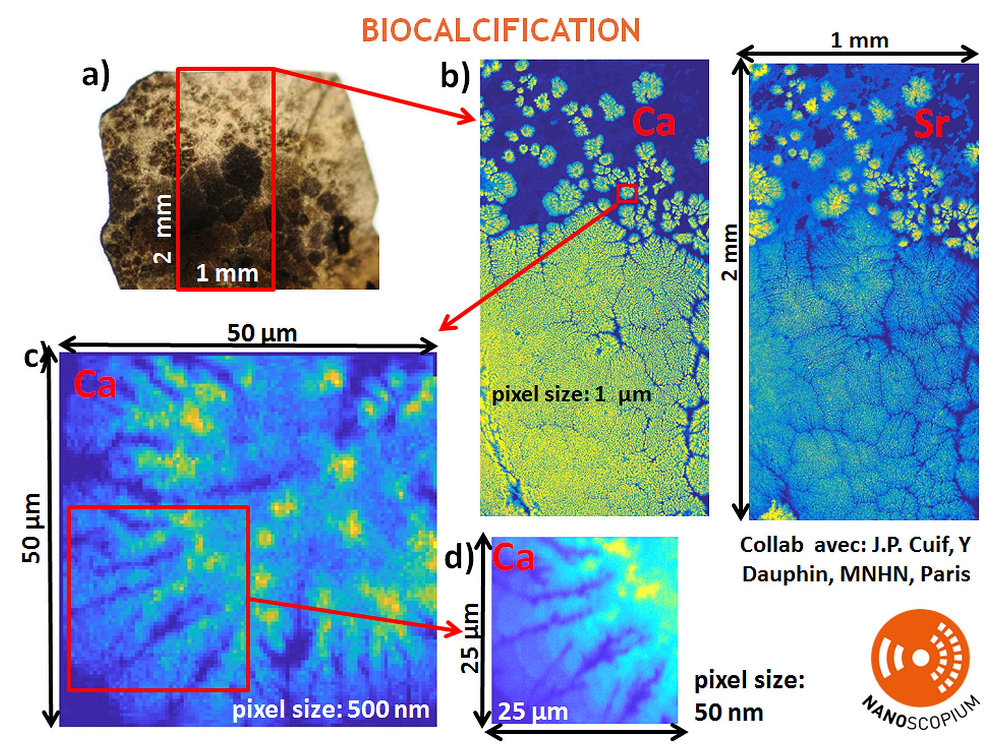
Calcium (Ca) and Strontium (Sr) distribution of the nacreous layer of a young pearl measured by scanning X-ray fluorescence microscopy. a) Optical microscopy image of the sample. b) Repartition of Calcium (Ca) and Strontium (Sr) within a 1 mm x 2 mm sample area with micrometer spatial resolution. c-d) "Zoom-in" of the Ca distribution within the regions of interest indicated by red rectangles in b and c. c) Region of interest chosen from b: size: 50 micron x 50 micron, spatial resolution: 500 nanometre, d) Region of interest chosen from c: size: 25 micron x 25 micron, spatial resolution: 50 nanometre.
Coherent Diffraction Imaging: down to 35 nanometre spatial resolution
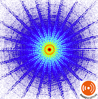
Sample: Siemens star, energy: 11.2 keV, exposure time: 1 s
Full field absorption and phase contrast X-ray micro-tomography: spatial resolution ~1.2 µm per voxel, acqusition time per tomogram ~5 minutes.
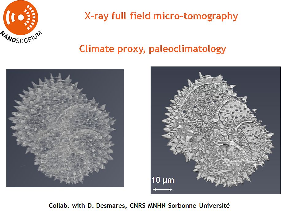
Volume rendering of the absorption contrast tomogram of a foraminifera sample. Full field X-ray micro-tomography techniques are available in combination with scanning multimodal imaging.
Under development: Scanning 2D/3D XRF-tomography and complementary modalities.
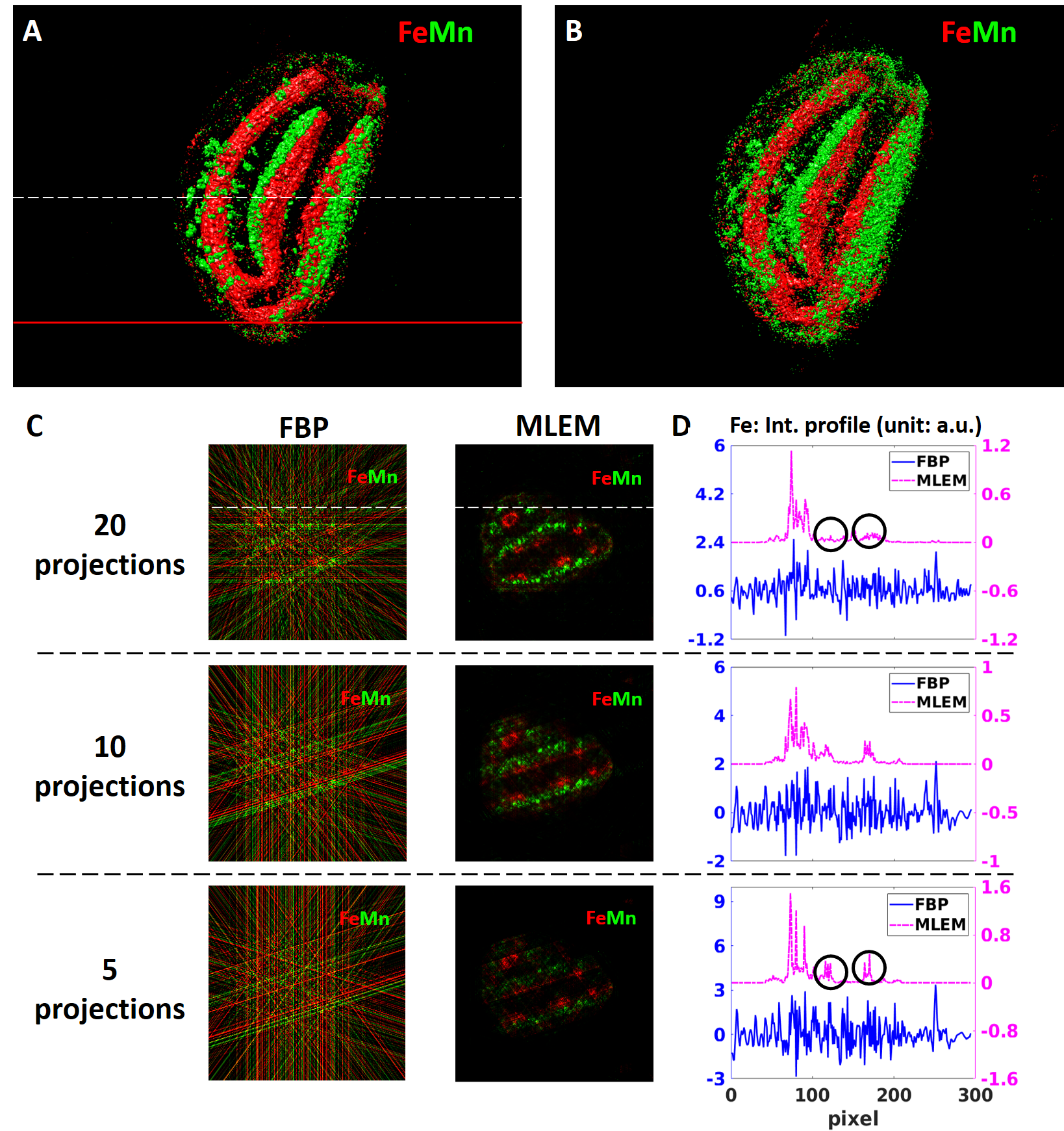
Our paper on non-invasive multi-length scale scanning XRF-tomography and complementary modalities is available in Scientific reports. The proposed robust, holistic workflow, including semi-automatic data reconstruction, opens the way towards routine multimodal 3D characterization of intact samples. Sci Rep 12, 16924 (2022). https://doi.org/10.1038/s41598-022-21368-0.
Scientific Opportunities
Earth Sciences & Geobiology | Biocalcification, micro-fossils, paleo-geochemistry, microorganisms in rocks and soils |
|---|---|
Environmantal Sciences | Pollution, bioremediation, climate proxies, paleoclimatology |
Biology-Health | Metals in cells/tissues: localisation, (mis-)regulation, accumulation, mineralization, biotechnology |
Material Science | Microelectronics, energy storage materials, functional devices, nano-structures, dopants, buried structures |
Cultural Heritage | Art, history, archaeology and conservation science, pigments, ceramics, resins, fibers. |
Contacts
Andrea Somogyi Proposals, Scientific & Methodological questions | Office: 01 69 35 96 46, Cell phone: 06 45 47 85 59 andrea.somogyi@synchrotron-soleil.fr |
|---|---|
Kadda Medjoubi Proposals, Scientific & Methodological questions | Office: 01 69 35 96 64 kadda.medjoubi@synchrotron-soleil.fr |
Gaëtan Correc Technical Affairs | Office: 01 69 35 94 42 gaetan.correc@synchrotron-soleil.fr |
Beamline News
All news of the Beamline
SOLEIL's contribution to the Energy Transition
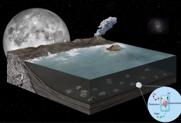
Paleontology: how did early life forms survive in an Arsenic-rich ocean?
Team

* External service provider, temporary or collaborator
Videos
All Nanoscopium's Videos
The lights of SOLEIL
Presentation of future beamlines Nanoscopium and Anatomix
Technical data
- Energy range
-
From 5 KeV to 20 KeV
- Energy Resolution
-
ΔE/E = 10-4 (Si 111)
- Source
-
Cryo-U18 undulator
- Flux @ first optical element
-
White beam
- Optics
-
Entrance Optics: Mirrors (vertical and horizontal focusing) for prefocusing and harmonics rejection (Si and Rh coating).
Monochromator: Fixed exit Double Crystal Si(111)
Nanofocusing optics:- CX3 station:High optical quality Kirkpatrick-Baez (KB) mirror-pair (JTEC), its opto-mechanical system has been provided by Bruker
- CX2 station:Fresnel Zone plates (FZP) developed in collaboration with PSI (Christian David, Laboratory for Micro- and Nanotechnology)
- https://www.synchrotron-soleil.fr/sites/default/files/endnote-pdf/mohacsi-2016-fabrication_and_characterization_.pdf
- https://www.synchrotron-soleil.fr/sites/default/files/endnote-pdf/mohacsi-2015-high_resolution_double-sided_diff.pdf
- Experimental techniques
-
Multi-technique and multi-lengthscale scanning X-ray imaging and tomography:
Analytical techniques:
- X-Ray Fluorescence (XRF) nanoprobe: open for user applications
- X-ray Absorption Spectroscopy (XANES): open for user applications
- Full field X-ray microtomography: open for user applications
- XRF tomography: please contact the beamline scientists
- Absorption-, Phase-, and Dark Field contrast imaging & tomography: please contact the beamline scientists
- Coherent Diffraction Imaging (Ptychography): please contact the beamline scientists
Acquisition modes:
- 2D at multiple lengthscales
- 3D by full field microtomography tomography & scanning tomography
- Fast continuous sample scanning (FLYSCAN, multi-detector architecture) with down to ms dwell time/pixel
- Network architecture: 10 Gbits tailored to the au high data flux produced by the ensemble of detectors (1 TOctets per day)
- Sample Environment
-
Two experimental end-stations, CX2 et CX3, are in exploitation :
CX2 :
- Nano-focusing optics: FZP
- Fast multimodal imaging with high spatial resolution (down to 35 nm by Ptychography)
CX3 :
- Nanofocusing optics: KB (JTEC)
- Fast multimodal imaging with high spatial resolution (70 nm) and high photon flux providing both very high analytical sensitivity and high resolution for elemental and chemical characterisation
- Spatial resolution at sample
-
CX2 end-station: ~100 x 100 nm2 to 1 x 1 µm2 by FLYSCAN, ~35 x 35 nm2 by Ptychography
CX3 end-station: ~50 x 50 nm2 to 1 x 1 µm2 by FLYSCAN and by step-scan
- Flux on sample
-
CX2 end-station: 108-9 ph/s at 15 keV
CX3 end-station: 1010 ph/s at 15 keV
- Detectors
-
Single element Si Drift Detector (SDD, Ketek)
Multi-element XRF (4 SDD) (RaySpec)
Fast digital multichannel analyser:
- 4-channel FALCON (Xia, inc)
Pixel-detector: EIGER 500K
Pixel-detector: JUNGFRAU 500k
2 X-Ray camera with inderect conversion:
- Scintillateurs + optics with magnifications of (G2, G5, G10) ORCA FLASH
- Scintillateur + optics with magnification of (G4) PCO.Edge
Proposed 2D &3D imaging modalities
| X-ray Fluorescence Microscopy | elemental composition (identification/quantification of all the elements between sulphur and uranium) |
|---|---|
| X-ray Absorption Spectroscopy | chemical speciation |
| Absorption, phase and dark field contrast | morphology (electron density) |
| Ptychography (coherent diffraction imaging) | high spatial resolution morphology (~35 nm) |