Paleontology: how did early life forms survive in an Arsenic-rich ocean?
How did the earliest life forms manage to endure the extreme conditions of their environment, such as chemically toxic surroundings? When did they begin to develop survival strategies?
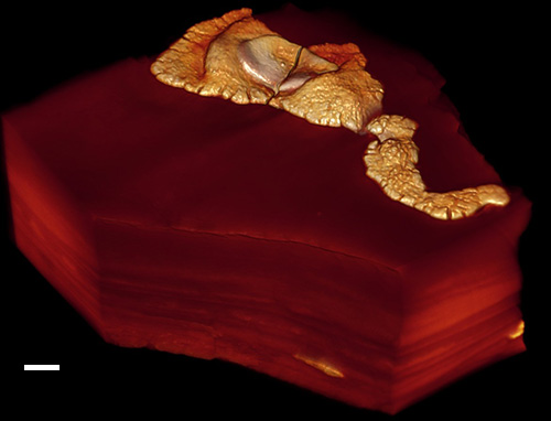
An international team of researchers led by Professor El Albani (IC2MP – CNRS / University of Poitiers) studied fossils from the Franceville Basin in Gabon, dated to 2.1 billion years ago, which include some of the oldest known eukaryotes¹. For the first time, the researchers identified arsenic confined within specific compartments of these fossilized organisms.
These results, obtained notably through nano–X-ray fluorescence spectroscopy on the NANOSCOPIUM beamline, suggest that these organisms developed a sequestration strategy to avoid arsenic poisoning. This kind of detoxification mechanism could represent one of the earliest direct pieces of evidence of environmental adaptation in the history of life.
Arsenic is a well-known toxic element, harmful to all known life on Earth. The study presented here suggests that some of the earliest complex life forms on our planet had already found ways to survive in the presence of arsenic. Indeed, the results show that these ancient organisms were capable of storing and isolating arsenic within specialized compartments inside their cells—an effective way to neutralize its toxicity.
To demonstrate this, scientists compared the presence and location of arsenic in fossils from the Franceville Basin (Figure 1) with that in more recent eukaryotic fossils (500 million years old) as well as in abiotic concretions. These fossils and concretions were chosen because the conditions—especially chemical ones—under which they formed are similar to those of the Francevillian fossils.
The X-ray fluorescence spectroscopy technique, carried out in collaboration with the team on the NANOSCOPIUM beamline, enabled the scientists to produce chemical maps at multiple length-scales and spatial resolutions. Being able to observe both an overview information on the variation of the elemental composition of these samples at a mesoscopic scale (several millimeters) and a zoom into deca-micron features with high resolution (deca-nanometers) was crucial to revealing the accumulation of arsenic and other elements (e.g., S, Ni, Fe, K, Cu) in micrometer-sized compartments within the analyzed fossils.
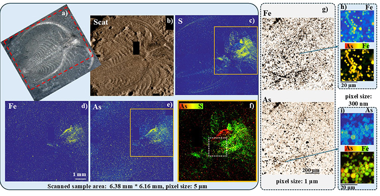
b. X-ray scattering signal (representative of the sample’s morphology),
c. sulfur distribution,
d. iron distribution,
e. arsenic distribution in the fossil at the mesoscale. f. RGB color representation showing colocalization of arsenic (As) and sulfur (S) in the head and stomach regions (highlighted in orange in images c and e).
g. Higher-resolution µ-XRF imaging (1 micron per pixel) in the intestinal area (outlined by a white dashed rectangle in f), revealing arsenic-rich pyrite microcrystals arranged in a filament-like network.
h–i. High-resolution (300 nm) distribution maps of iron and arsenic, showing that arsenic is unevenly distributed within the micropyrite crystals.
These insights into the elemental distribution in fossilized eukaryotes and abiotic concretions allowed the scientists to conclude that the arsenic enrichment observed in the 2.1-billion-year-old organisms was not the result of later contamination but instead reflects a biological response to environmental stress. They observed that the arsenic patterns in these fossils are distinct from those in inert mineral structures, strengthening the case that these were indeed complex, organized living organisms. This biological innovation was not random but closely linked to environmental changes, notably the rise in atmospheric oxygen around two billion years ago.
This research involved scientists from the NANOSCOPIUM beamline at SOLEIL, as well as from the universities of Paris-Saclay, Toulouse, Cardiff (UK), Lausanne (Switzerland), and the Natural History Museum in Vienna (Austria).
---------------------------------
1 – Eukaryotes: organisms —unicellular or multicellular— whose cells contain a nucleus and organelles (such as mitochondria) enclosed within membranes. In contrast, prokaryotic cells —like those of bacteria— have neither a nucleus nor organelles.