
Visualizing how an adjuvant boosts the effectiveness of antibiotics against a resistant bacterium
A collaborative study between Aix-Marseille Université, SOLEIL, ESRF and ALBA synchrotrons, recently published in npj Antimicrobials and Resistance, provides new insight into the mechanism of action of NV716, an antibiotic adjuvant capable of restoring the activity of certain antibiotics against the multidrug-resistant bacterium Pseudomonas aeruginosa.
Gram-negative bacteria such as P. aeruginosa exhibit strong intrinsic resistance due to the low permeability of their outer membrane and the efficiency of their antibiotic efflux systems. Certain adjuvants, such as NV716, enhance the effectiveness of antibiotics against these resistant bacteria. But how do they work?
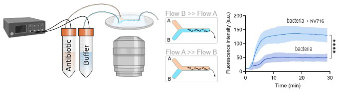
To understand the mechanisms involved, several complementary synchrotron imaging approaches were combined. At the DISCO beamline of Synchrotron SOLEIL, deep ultraviolet (DUV) microspectrofluorimetry experiments enabled real-time monitoring of antibiotic accumulation (DUV-fluorescent antibiotics) in individual bacteria using a dedicated microfluidic device developed for the study (Fig. 1). This device greatly improves the monitoring of antibiotic accumulation in bacteria using UV microscopy, by allowing to record the initial stages of accumulation and to overcome the issue of bacterial mobility before and during the uptake process.
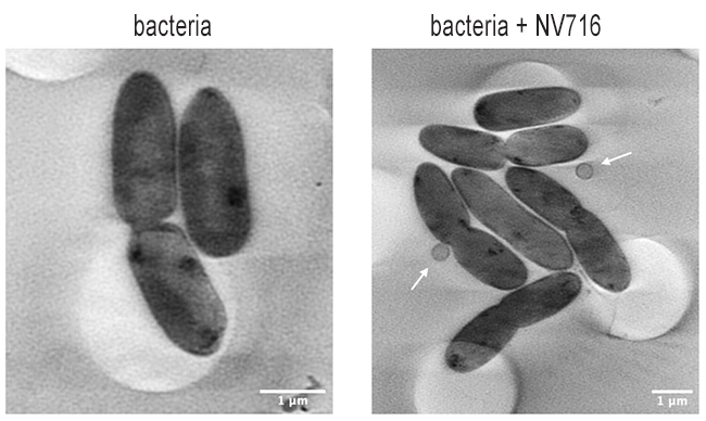
Cryo-soft X-ray tomography analyses performed at the MISTRAL beamline of the ALBA Synchrotron then revealed membrane alterations in bacteria exposed to the adjuvant, associated with increased production of outer membrane vesicles (OMVs) (Fig. 2).
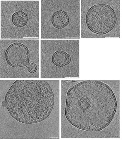
These observations were complemented by cryo-electron microscopy experiments carried out at SOLEIL (POLARIS platform), enabling visualization of the ultrastructure of vesicles produced in the presence of NV716 (Fig. 3).
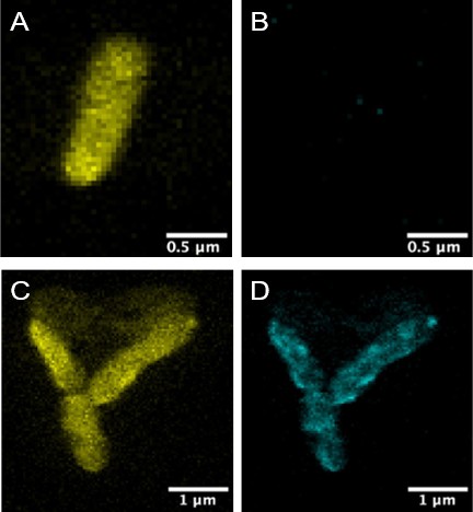
Finally, X-ray fluorescence nano-imaging experiments conducted at the ID16A beamline of the ESRF enabled localization of a copper-containing derivative of the adjuvant (Cu-NV716) at the periphery of the bacteria, confirming its association with bacterial membranes (Fig. 4).
These results show that NV716 acts primarily by disrupting the organization of the outer membrane, thereby promoting intracellular accumulation of antibiotics such as doxycycline.
Beyond elucidating the mode of action of NV716, this work opens new perspectives for exploring the mechanisms of OMV biogenesis and for developing strategies aimed at modulating membrane permeability in multidrug-resistant bacteria.