Paleontology: How microscopic plankton played a major role in the formation of limestone in the Cretaceous seas
A collaborative team of micropaleontologists, geochemists, and physicists from ISTerre (CNRS/Université Grenoble Alpes), CEREGE, Institut Néel, SOLEIL, and Rutgers University provides new insights into the mechanisms of skeletal formation in Nannoconus, an extinct calcareous microplankton that played a major role in biocalcification in the Cretaceous seas.
For nearly 35 million years, the exoskeletons of this genus have contributed to massive carbonate accumulations on the seafloor, potentially impacting seawater chemistry. Despite their geohistorical importance, the fine-scale skeletal organization and the associated calcification processes have remained largely unresolved.
The Nannoconus cone-shaped exoskeleton consists of an assemblage of micaliths, themselves composed of imbricated calcitic components, but until now their microstructure was not resolved at scales relevant for discussing biomineralization processes. By combining X-ray ptychographic computed tomography (PXCT) on the SWING beamline at the SOLEIL synchrotron with scanning electron microscopy, this study provides the first three-dimensional reconstruction of a Nannoconus micalith at nanometric resolution, below the thickness of its constituent lamellae.
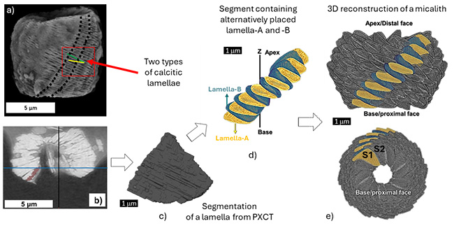
The results reveal a highly hierarchical organization: each micalith is composed of segments formed by the spiral stacking of calcite lamellae with two alternating inclinations, and building a wall around a central canal. This geometry imposes strong constraints on mineralization modalities and supports the hypothesis of biologically controlled mineralization, involving organic matrices in the orientation and arrangement of the lamellae.
This first characterization opens the way for comparative analyses at the generic level, and subsequently at the ordinal level of the Braarudosphaerale to which Nannoconus belongs, in order to assess the role of morphological selection throughout their long evolutionary history.
It also highlights the relevance of these crystalline microarchitectures as natural analogous for the development of biomimetic materials whose functional properties are governed by multiscale organization, particularly in catalysis and biomedicine.