
Malaria: a new target identified for blocking liver infection using antibodies
Malaria parasites must first infect the liver before they can cause disease. Scientists from the Laboratory of Medical Biochemistry (University of Antwerp) and the Centre d’immunologie et des maladies infectieuses (Sorbonne Université, Inserm, CNRS) combined structural modelling, electron microscopy, small-angle X-ray scattering at the SOLEIL beamline and antibody neutralization assays to investigate two key Plasmodium sporozoite proteins, P36 and P52.
Their results suggest that P36 and P52 form a complex that can be targeted by neutralizing antibodies, preventing infection of liver cells. These proteins could therefore represent promising targets for new antimalarial strategies.
Malaria is caused by parasites of the genus Plasmodium, which are transmitted through the bite of infected Anopheles mosquitoes that inject parasite stages known as sporozoites (SPZs) into the host skin. SPZs migrate to and infect the host liver for an initial round of multiplication. SPZs and liver stages have long been considered as ideal targets for malaria vaccines as targeting these stages can prevent disease onset.
During the parasite’s life cycle a multitude of stage-specific surface proteins are involved in a variety of important processes. The main SPZ surface protein, the circumsporozoite protein (CSP), is the target of currently approved malaria vaccines and preventive antibody therapies. Studies in mouse malaria models have shown that antibodies against CSP protect mainly by stopping the parasite in the skin. However, some of the most effective antibodies also provide protection against the parasite in the bloodstream and liver. Other SPZ proteins involved at different steps of the infection process may thus represent additional targets for protection.
Three members of the 6-cysteine protein family P36, P52 and B9 play an essential role during SPZ invasion of liver cells, yet their function and whether they can be targeted by neutralizing antibodies remain unknown. Here, to investigate these unanswered questions, the scientists combined advanced structural biology techniques with functional experiments in a malaria mouse model.
Artificial intelligence-based protein structure prediction and experimental structural data obtained through electron microscopy (see figure 1) and small-angle X-ray scattering (SAXS, performed at the SOLEIL SWING beamline, see figure 2), suggest that P36 and P52 interact to form a heterodimer.
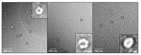
This heterodimer displays a head-to-tail orientation with a protein-protein contact region that is largely maintained across different Plasmodium species.
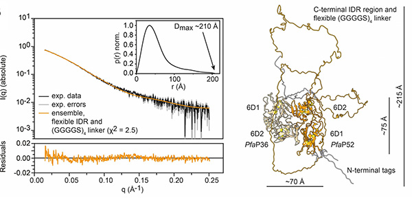
The structural models of the heterodimer guided the design of experiments in which epitope tags were added to P36-P52 heterodimers that were either located proximally or distally with regards to the parasite surface. Both types of tagged heterodimers were subsequently subjected to anti-tag antibodies. Neutralization assays revealed that, in culture conditions, antibodies against membrane-distal tagged P36 and P52 can efficiently block invasion of liver cells (see figure 3).
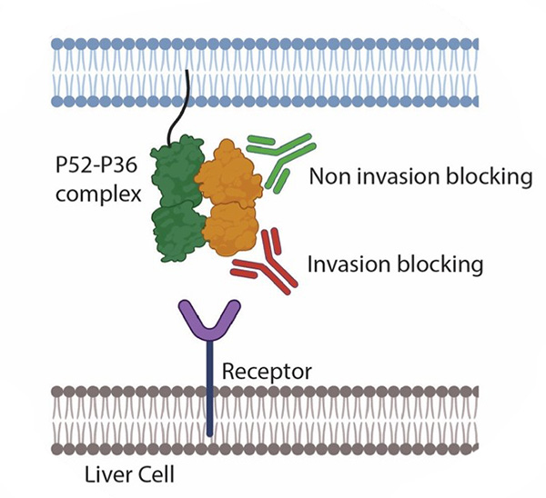
This data shows that the inhibitory activity of anti-tag antibodies heavily depends on the tag position and revealed that antibody-exposed vulnerable sites lie on the membrane-distal side of the P36-P52 complex. In contrast, antibodies against tagged B9 had no inhibitory effect on SPZ invasion, irrespective of tag positioning.
Although many questions remain about the structure-function relationship of P36 and P52 (for which a high-resolution structure would be of great value), these data show that the liver invasion step could be targeted by antibodies and indicate that the P36-P52 complex might be considered as a potential target for the development of new liver stage malaria vaccines or therapeutic antibodies.