Deciphering DNA and RNA 3D structures using circular dichroism
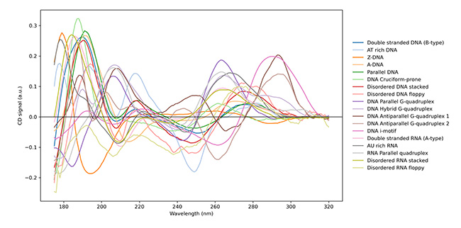
NA-CDQuant is a novel deconvolution algorithm which transforms Circular Dichroism CD spectroscopy from a qualitative to a quantitative method for nucleic acid analysis. Building on the NACDDB nucleic acid CD/SRCD database, NA-CDQuant defines 18 reference spectra to distinguish overlapping spectral features and identify DNA and RNA conformations in solution.
This tool provides a valuable approach for assessing the formulation and quality control of nucleic acid-based therapeutics. The project was developed through a collaboration between DISCO beamline, CEA/LLB, SDSM&T (USA), ISA (DK) and Sanofi.
Nucleic acids (NAs) are no longer viewed solely as passive carriers of genetic information. DNA and RNA adopt a variety of structural conformations in solution, ranging from canonical double helices to motifs such as quadruplexes or pseudoknots. These structures govern essential biological processes including gene regulation or catalytic activity (ribozymes). These structural arrangements are highly sensitive to their environment and subtle shifts in ion concentration, temperature, or molecular crowding can alter thermodynamics equilibria and consequently, NAs biological functions. This structural plasticity is a cornerstone of cell biology and underpins the rapidly expanding field of NA–based medicines, such as therapeutics using coding and noncoding RNAs.
While high-resolution techniques such as X-ray crystallography, NMR and cryo-electron microscopy provide detailed structural insights, they are often constrained by requirements for sample crystallinity, high concentrations, or specific molecular sizes. In contrast, optical spectroscopies offer a rapid and non-destructive alternative for monitoring folding and environmental responses in solution without size limitations. Among these, circular dichroism (CD) spectroscopy is particularly powerful due to its sensitivity to the chiral arrangement of NAs. In the ultraviolet range, CD signals arise from electronic transitions, producing characteristic spectral patterns that reflect helicity, base stacking, pairing, and overall secondary structure.
Despite its potential, the application of CD spectroscopy to NA structure analysis has remained largely qualitative. While quantitative interpretation methods are well-established for protein characterization, NAs present a unique challenge: they populate heterogeneous ensembles of conformations. Canonical helices, partially folded intermediates, and noncanonical structures often coexist in a sample, producing composite, overlapping spectra that are notoriously difficult to interpret. Furthermore, while Synchrotron Radiation Circular Dichroism (SRCD) has extended the accessible wavelength range and increased information content, the field lacks a standardized, comprehensive references framework. This absence has limited CD analysis to simple "fingerprinting," leaving a critical gap in the ability to perform the precise, quantitative structural assessments required for modern drug development and quality control.
To address this challenge, NA-CDQuant is a novel deconvolution algorithm designed to transform CD spectroscopy from a qualitative descriptive tool into a precise quantitative analytical method. By integrating more than 200 UV SRCD spectra deposited in the nucleic acid spectra CD database (NACDDB), the algorithm is based on 18 distinct reference spectra to define specific conformational families. This systematic approach overcomes the long-standing obstacles of spectral overlap and structural polymorphism, enabling the accurate determination of the fractional composition of coexisting DNA or RNA conformations in real-time. The development of NA-CDQuant thus provides a practical bridge between NA molecular spectroscopy and functional biological insights. By facilitating precise assessment, this framework serves as an essential tool for the development of emerging NA–based therapeutics. This breakthrough is the result of an extensive international collaboration between DISCO beamline at Synchrotron SOLEIL, CEA/LLB, SDSM&T (USA), ISA (DK) and Sanofi.