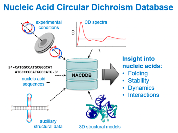
NACDDB: Nucleic Acid Circular Dichroism DataBase
A novel database has been created in a concerted scientific approach. Researchers from Poland, France and the United States have joined efforts to collect and standardize Circular Dichroism (CD) spectra of nucleic acids, DNA or RNA. More than 150 spectra, taken from the literature or recently collected at the DISCO beamline, have been gathered in the “NACDDB” and made accessible to the public via a webserver (https://genesilico.pl/nacddb/).
Nucleic acids such as DNA, known for harboring the genetic information in form of chromosomes, or mRNA, recently used in antiviral treatment (COVID-19), are macromolecules made of a phosphate backbone, sugar (oxy or deoxy ribose) and 5 different nitrogen-containing molecules called bases (purines and pyrimidines, called A, C, G, T and U). Their molecular structure or spatial arrangement, such as the double stranded helix of DNA, is crucial for their function.
Circular Dichroism spectroscopy measures the differential absorption of circular right and left polarized light in the Ultra-Violet range by chiral* molecules, like nucleic acids for instance. In the spectral range between 7 and 5 eV, the electrons of the nucleic acid macromolecules are excited by the light and are at the origin of the UV absorption. Characteristic absorption bands of circular (right or left) polarized UV light (commonly referred to as UV -A-B-C and specifically between 320 -170 nm) allow to distinguish different types of folding of these complex macromolecules. Their spatial arrangement correlates to the CD spectra and ultimately aids to determine the folding of nucleic acids.
For the past two decades structural biologists have collected, deposited and screened CD spectra obtained from proteins permitting them to successfully determine secondary structure content (alpha helices, beta sheets and random coils) for amyloids, membrane proteins and protein-protein complexes notably.
The difference between nucleic acids and proteins consists in the chiral molecule also called chromophore: for proteins the main chromophore is the peptide bond linking covalently the amino acids, which is small in size. In the case of nucleic acids, the chromophore is a nucleotide consisting of a phosphate, ribose and a base. It is much larger and variable in size compared to peptide bond. The sheer multitude of distances observed, e.g. single stranded or double stranded nucleic acids, leaves a lot of possibilities for the attribution of electronic excitations.
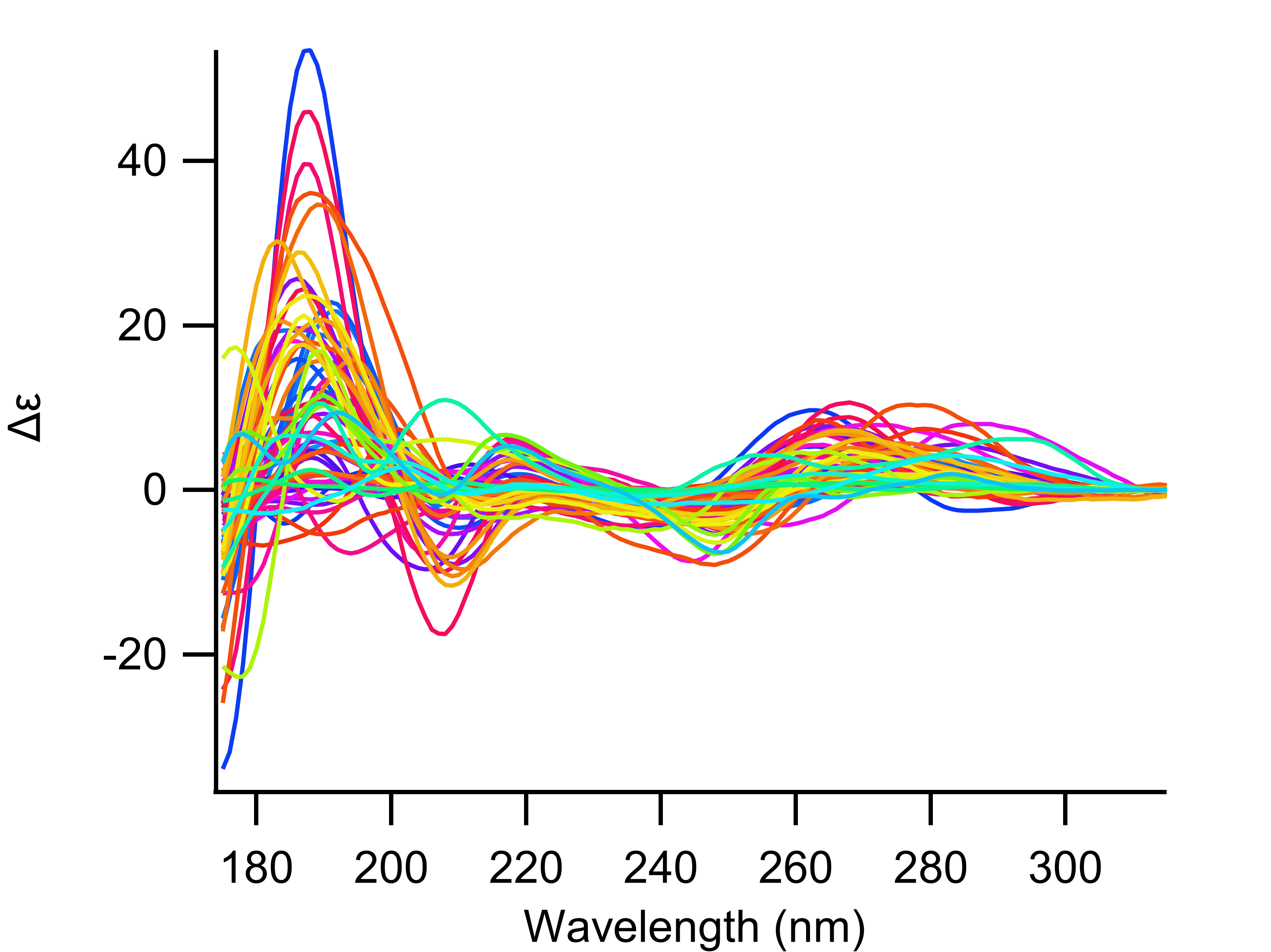
Keeping this in mind and searching the literature, scientists have found published spectra dating back as far as the late 50’s. Their first goal has been achieved now by systematically digitalizing nucleic acid CD spectra from the literature. In addition, data have been added collected recently at the DISCO beamline at SOLEIL, 63 Synchrotron Radiation Circular Dichroism (SRCD) spectra have been collected from common and special (e.g. methylated nucleotides) DNA and RNA macromolecules in solution (figure 1), extending the information content accessible in addition to existing top bench CD spectra from the literature.
All these spectra (more than 150) have been deposited in the NACDDB public repository, which archives and freely distributes the experimental data including experimental metadata (experimental conditions like pH, salinity and temperature), structural models and links to the corresponding references. Various types of nucleic acid molecules, including DNA, RNA, complexes of the two, and -last but not least- modified nucleic acid derivatives, which are of pharmaceutical interest, have been deposited.
The database allows for searching, comparing, downloading and in future will allow to analyse CD spectra for nucleic acid structural arrangements. For known sequences with related high-resolution structures, 3D models are available too. The NACDDB is also open for submission of CD data from nucleic acids taken in structural-biology laboratories on top-bench spectrophotometers as well as on synchrotron radiation facilities providing SRCD.
*chiral (greek word for 'hand'), referring to anything which can’t be superimposed on its own mirror image. Chiral molecules contain chiral centers like the tetrahedral carbons with four different substituents (like the carbon atoms involved in the peptide bonds of proteins).