
POLARIS
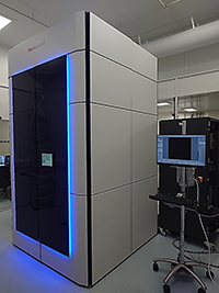
The Titan KRIOS G4 transmission cryo-electron microscope (Cryo-EM) at SOLEIL, POLARIS, was financed by the French government as part of the EQUIPEX France-Cryo-EM (Contract ANR-21-ESRE-0046) and is operated as a user facility by Synchrotron SOLEIL.
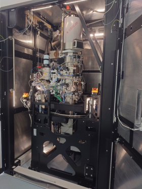
Operational since October 2024, POLARIS delivers a high brilliance, coherent source of up to 300 keV electrons to cryogenically vitrified biological samples prepared on AutoGrids. The samples can be distributed solutions of biological macromolecules or their complexes for Single Particle Analysis (SPA), or thin samples (viruses, vesicles) or sections of either frozen cells or biological tissue samples for Cryo-Electron Tomography (Cryo-ET). Equipped with a cold Field Emission Gun (FEG), an energy filter and a rapid and sensitive direct electron detection camera (Falcon4i), POLARIS can produce high resolution (from 1 – 5 Å resolution) images of biological systems for both pure and applied research in biological science, thus contributing to the palette of techniques available in the Life Sciences scientific section at SOLEIL. The Cryo-EM measurements are highly complementary to X-ray crystallographic and Small Angle X-ray Scattering measurements performed on PROXIMA-1, PROXIMA-2A and SWING beamlines respectively.
Team / Contact
POLARIS Phone number: 01 69 97 59
Members
Facility Manager
Pierre Legrand
01 69 35 96 60
06 86 42 32 97
Facility Operation Manager
Éric Larquet
01 69 35 96 00
06 78 10 61 53
Opérator
Heddy Soufari
01 69 35 96 69
Technical data
- Model
-
Thermo Fisher Scientific KRIOS-G4
- Electron source
-
Cold FEG which can operate at 300 kV or 200 kV
- Detection
-
Falcon 4i (up to 320 frames per second readout speed in EER).
File format: EER format, or TIFF.
- Energy filter
-
Selectris X
- Sample stage
-
X-Y positioning and single axis rotation range from -70 - + 70 degrees
- Additional information
-
The microscope is fitted with a Volta Phase plate, and an autoloader capable of storing up to 12 Cryo-EM auto-grids at cryogenic temperatures.
The POLARIS facility can produce very-high to high resolution 3D images of biological systems.
In Single Particle Analysis mode, a vitrified solution of intact particles in random orientations is imaged with achievable resolution up to 1.1 Å in highly favourable cases but with 2.0 – 3.5 Å structures routinely achievable depending on the sample type. Automation of image processing steps is achieved by the SCIPION3 interface (Biological Imaging , Volume 3 , 2023 , e13 DOI: https://doi.org/10.1017/S2633903X23000132) or Cryosparc live (for academic users). A typical SPA experiment consists of microscope pre-alignment by the facility team, introduction and inspection of Cryo-EM grids and selection of grid co-ordinates to be collected, following by automatic data collection of images. A typical experiment takes 1 – 2 days of measurement time. After data collection, raw and pre-analysed data can be backed up onto external USB disk or recovered by the GLOBUS application. Data can also be pushed to AWS storage or rsynced.
For further information on sample preparation, please contact the operations team.
In Cryo-Electron Tomography Mode, lamella cut from vitrified cells or tissues can be measured by POLARIS using the sample tilt stage to acquire a dose-symmetric tilt sequence of images from which tomographic reconstruction of the sample can be performed for example by using back-projection algorithms.
For further information on sample preparation, please contact the operations team.
Access
Applications for access can be made via the SunSET, selecting the Rolling Access or Block Allocation group (BAG) modality. In Rolling Access, proposals can be submitted at any time, but will be cumulated and reviewed every 2 months. Consequently the time between project submission and attribution of access will be up to 3 months. See more details here.
Access to POLARIS is free of charge to academic projects or industrial-academic partnerships approved by the SOLEIL Programme Committee for biology and health, and for which results will be published in a peer-reviewed journal. For proprietary access to the microscope, users should contact the industrial outreach group for advice and competitive quotation.
Sample Handling
Users are asked to bring or send pre-clipped AutoGrids – (exceptionally, the clipping of the grids can be performed on-site) and their introduction to the microscope will be performed by in-house staff in order to optimise autoloader reliability. Dry-shipping Dewars can be sent to
Synchrotron SOLEIL
L’Orme des Merisiers
Départementale 128
91190 Saint-Aubin
The shipment should be clearly marked with the name of your local contact for the experiment, and tracking numbers should be provided to enable the dry-shipper to be refilled on arrival.
Assistance can be provided in sample preparation for a small number of inexperienced users. SOLEIL is equipped with both Thermo Fisher Vitrobot and LEICA EM GP Cryo-plunge systems.
Data Storage and Analysis
Data will be stored on site according to the principles laid out in SOLEIL’s data management policy, for at least a 5 years. Longer term data storage cannot be guaranteed.
Although processing and analysis of Cryo-EM data is complex and involves several passes through the data, POLARIS is providing a live processing (and is working at implementing the SCIPION3 pipeline for pre-data processing) which runs during the measurements and will give an initial idea of the quality of data being collected.