PSICHÉ
PSICHÉ (Pressure Structure and Imaging by Contrast at High Energy): this beamline is dedicated to x-ray diffraction under extreme conditions (pressure-temperature) and to tomography at high energy (20-120 keV).
The PSICHÉ (Pressure Structure and Imaging by Contrast at High Energy) beamline is dedicated to X-ray diffraction under extreme conditions (pressure-temperature) and to tomography at high energy, from ambient to extreme conditions. The source is an in-vacuum wiggler that is used to produce a wide variety of beam modes, from a large parallel white/pink beam to a microfocused monochromatic beam. PSICHE is the highest energy beamline of SOLEIL. It has two hutches, one dedicated to white/pink beam (energy range: 15-100+ keV) and another to monochromatic beam (20-50 keV). Extreme pressure and temperature conditions can be generated using different equipment: a 1200-ton multi-anvil press, Paris-Edinburgh presses (including tomography at extreme conditions), and diamond anvil cells (including a double-sided laser heating facility with 4 color pyrometry). The use of multiple techniques, and the combination of complementary techniques, are important features of the beamline.
The beamline uses 4 different beam modes:
- White/pink beam, parallel
- White/pink beam, focused (vertical only)
- Monochromatic beam, focused or microfocused
- Monochromatic beam, parallel
The three associated experimental stations are:
- CX1: white/pink beam (high-speed imaging, XRD, large volume presses)
- CX2: monochromatic beam, focused (vertical) or parallel (imaging and XRD)
- CX3: monochromatic beam, microfocused (micro XRD, LHDAC)
Beamline news
All PSICHÉ newsSUCCESS STORY – NEPHEWS Twinning Program – Ali Güzel, hosted at the PSICHE beamline from 3 (...)
World-first at SOLEIL: 3D X-ray imaging of pressurized hydrogen-induced damage in steel (...)
Team

* External service provider, temporary or collaborator
Videos
All PSICHÉ videos
A talisman against Yazdanduk
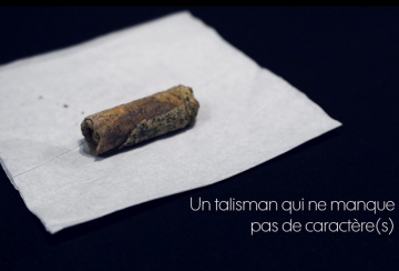
A talisman with plenty of character(s)
Technical data
- Energy range
-
15-100 keV for white beam (low energy part is filtered)
15-50 keV for monochromatic beam
25-120 keV for pink beam imaging
- Energy Resolution (ΔE/E)
-
∆E/E ~10-3 for monochromatic beam modes (Double Crystal Monochromator)
∆E/E ~10-2 for energy dispersive diffraction (Ge detector resolution)
∆E/E ~10-1 for pink beam imaging modes
- Source
-
Under vacuum multipole wiggler: 2.1 T, period 50 mm, 38 periods
- Optics
-
- Focusing vertical mirror
- Double Crystal Monochromator (Si111)
- Two focusing mirrors in KB geometry
- Multiple pink beam filter configurations
- Sample Environment
-
- Diamond anvil cells
- Paris-Edinburgh cell (4 column and UToPEC versions)
- Large Volume multianvil cell (1200 t, DIA module)
- Tomography furnace (<1400 °C, collaboration 3DMagination/Centre des Materiaux)
- Flux on sample
-
- White beam :1-8 108 ph/s/0.1% E in a 10 μm x10 μm hole
- DCM 111: at 30 keV 1.4 1013 ph/s in a 16.8 x 5.9 mm2 spot, 1.8 1012 ph/s in a 12 μm x10 μm spot after KB
- Pink beam: >2 1015 ph/mm2/s in a 11 x 3 mm2 beam
- Detectors
-
- Pilatus CdTe 2M
- Perkin-Elmer (now Varex) flat panel
- Tomography detectors
-
Lens coupled scintillator detectors, based on the following cameras:
- Hamamatsu ORCA Flash4.0 v2 and v3 for imaging
- Hamamatsu ORCA Lightning 4602 x 2596 pixels
- PCO Dimax HS4 (loan from ANATOMIX beamline)Pixel sizes from 0.13 to 10 microns.
Scientific Opportunities
| X-ray diffraction: | Materials under extreme conditions, Geosciences (planetary interiors), Physics (molecular solids, functional materials, highly correlated electrons materials…), chemistry (synthesis of hard material) Biology (folding and unfolding of proteins), ... |
|---|---|
| Tomography | Materials science, in-situ experiments including under high pressure, cultural heritage, biology and health, ... |
Beamline Layout

PSICHE beamline beam modes and corresponding experimental stations. a) Monochromatic beam, focused or microfocused b) White/pink beam, focused (vertical only) c) White/pink beam, parallel d) Monochromatic beam, parallel
The UToPEC (Ultrafast Tomography Paris – Edinburgh Cell) for structure, imaging, density and sound velocity measurements up to 15 GPa – 2000 K
The UToPEC is a new Paris-Edinburgh press optimized for fast tomographic imaging at high pressures and temperatures at the PSICHE beam line of the SOLEIL Synchrotron. This unique development means that previously inaccessible industrial or geological processes can be studied using synchrotron x-ray tomography with a time resolution of 1 second and a spatial resolution of a few microns for processes up to 15 GPa pressure and 2000K temperature.
Techniques compatible in a single experiment with the UToPEC are:
- Energy-Dispersive X-Ray Diffraction (ED-XRD) that can be extended to combined angle and energy (CAESAR)
- Fast X-ray Computed Tomography (XCT) in pink beam mode
- Beer-Lambert absorption method for density measurements (liquid, glasses, solid)
- Ultrasonic measurements
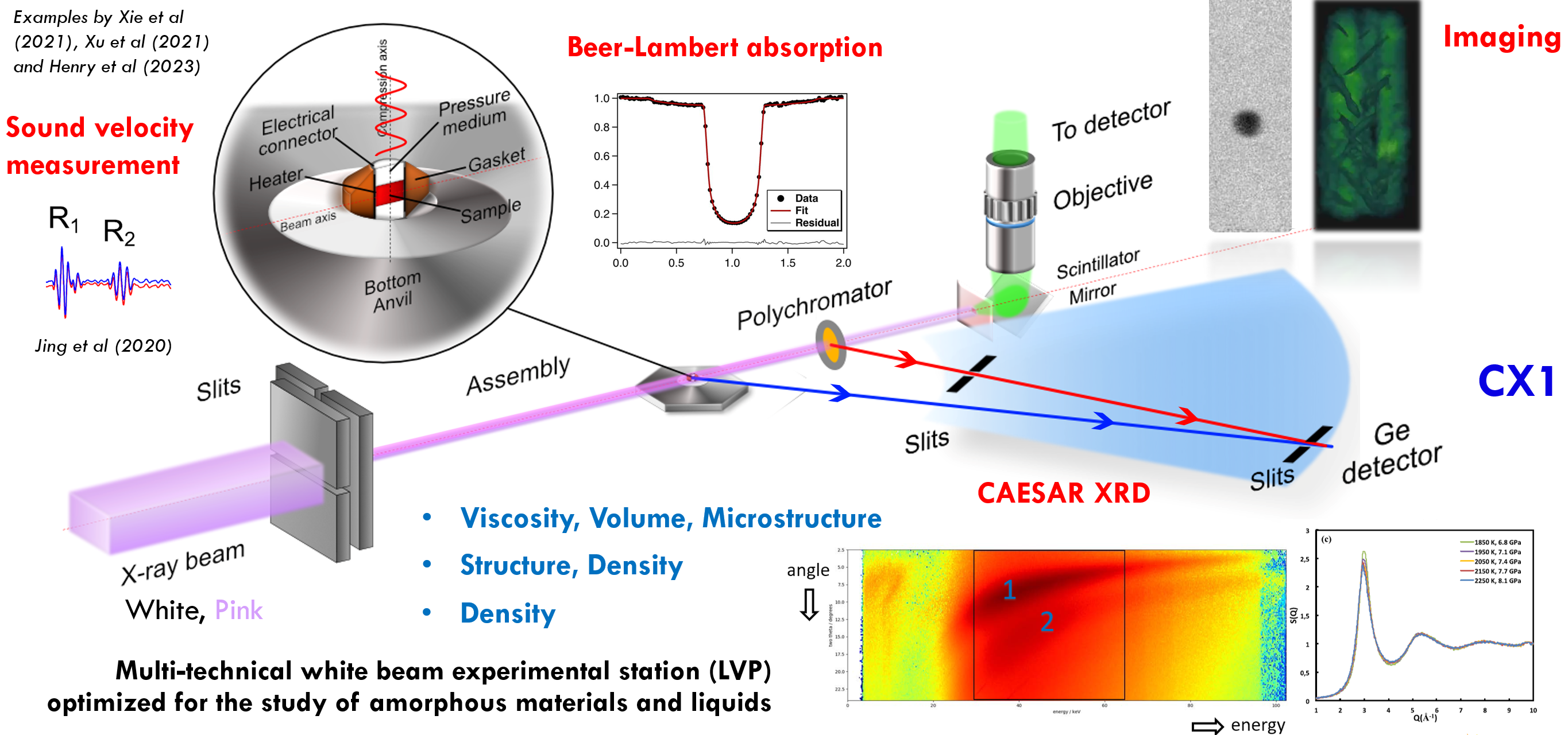
Schematic of the suite of techniques available at PSICHÉ using the UToPEC in white beam/pink beam mode (65 keV - tunable). (Credits: Nicolas Guignot)
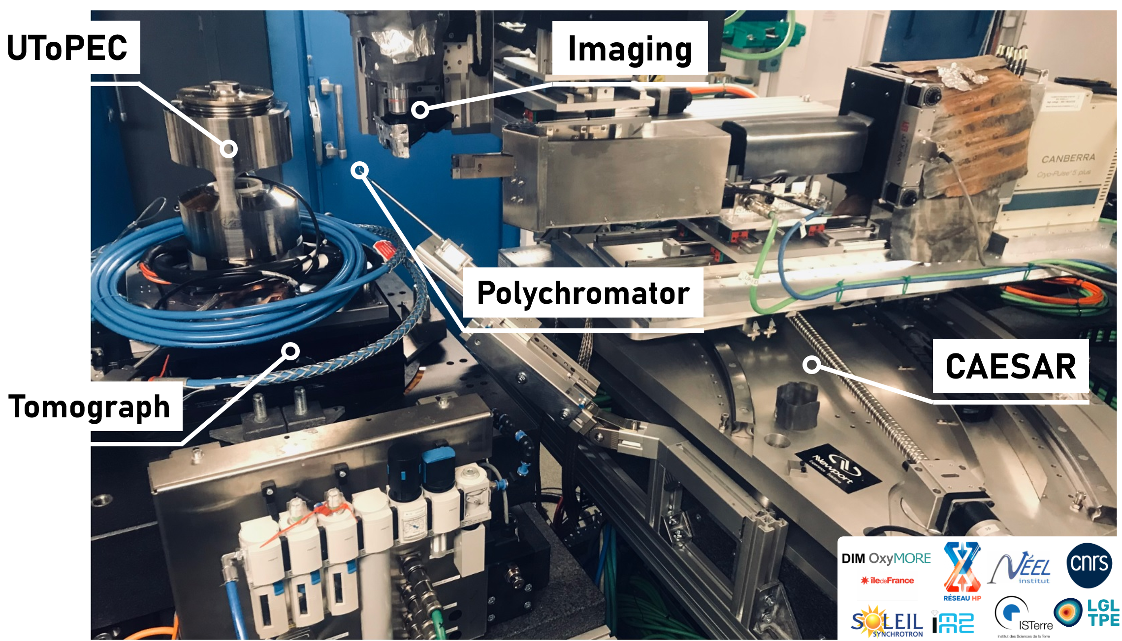
The UToPEC experimental station
An analysis package for data analysis and in-house software development can be found at: https://github.com/EarthianOnEARTH/analysis-package/
Contact: laura.henry@synchrotron-soleil.fr
Tomography (pink beam or monochromatic) accommodating various sample environments
PSICHE offers parallel beam microtomography and radiography which can be used for a wide range of applications. The instrument is highly flexible and can be adapted to different experiments: Materials science, biology, paleontology and cultural heritage, etc. Some important features are:
- Very large diameter rotation stage with a 250mm free aperture which can be used for installing large samples or heavy/bulky in-situ equipment.
- Spatial resolution as high as ~0.8 microns.
- Photon energies up to >100 keV.
- Tomography as fast as 0.5 seconds per complete volume.
- Field of view up to 11 mm x 3 mm (h x v).
- Pink beam or monochromatic beam modes. Pink beam mode allows fast or high energy imaging. Monochromatic is safer for delicate samples and enables K-edge subtraction imaging.
- Multi-resolution (“zoom in”) tomography.
- Tomography can be combined with diffraction in both pink beam and monochromatic beam modes. For example, to study both morphological and crystallographic phase changes during reactions, or in an operando battery.
- Fast and automated tomography reconstructions to follow your experiment in near real time.
Beamline references:
https://pubs.aip.org/aip/rsi/article/87/9/093704/365750
https://link.springer.com/article/10.1007/s40192-019-00155-2
Beamline code:
https://gitlab.com/soleil-psiche
Contact: andrew.king@synchrotron-soleil.fr
Laser-heated diamond anvil cell – Temperature mapping
Laser-heated diamond anvil cell (LHDAC) setups are designed to reach the highest P and T conditions using static compression methods. The PSICHE beamline setup stands out thanks to several features:
- Laser beamshaping: the lasers focal spots can be easily shaped, giving access to a wide array of power distributions, including flat-top, donut, etc.
- 4-color pyrometry: two 4-color pyrometers specifically designed for synchrotron applications are available, giving access to real-time T and emissivity mapping thanks to our in-house software. The combination with laser beamshaping is particularly effective.
- High resolution, high NA objectives: imaging and temperature measurements are made possible thanks to two high-end Schwarzschild objectives, developed in-house.
- X-ray transparent mirrors: we use a high flatness glassy carbon mirror downstream to ensure observations and measurements at high spatial resolution without significantly impacting XRD data.
Accessible wavelength is currently limited to 1070 nm (on both sides). Typical T range is 1000-4000 K.
XRD can be recorded in-situ thanks to our DECTRIS Pilatus 3 CdTe 2M detector. Typical exposure times are 2-5 seconds but can be lowered depending on the samples. The X-ray beam size is 10x12 microns (VxH).
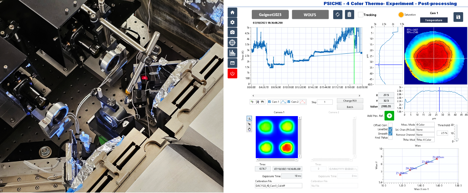
Left – LHDAC setup. Right - Snapshot of PSICHE’s 4-color pyrometry data recording and processing software.
Contact: nicolas.guignot@synchrotron-soleil.fr
The Multi-Anvil Press
The beamline is equipped with a compact, light and removable multi-anvil press with a DIA module. It has a 1200 tons capacity and 24° horizontal opening.
Techniques compatible in a single experiment with the multi anvil press are:
- Energy-Dispersive X-Ray Diffraction (ED-XRD) that can be extended to combined angle and energy (CAESAR)
- Fast X-ray Imaging (radiography) in pink beam mode for viscosity measurements (liquids)
- Beer-Lambert absorption method for density measurements (liquid, glasses, solid)
- Ultrasonic measurements
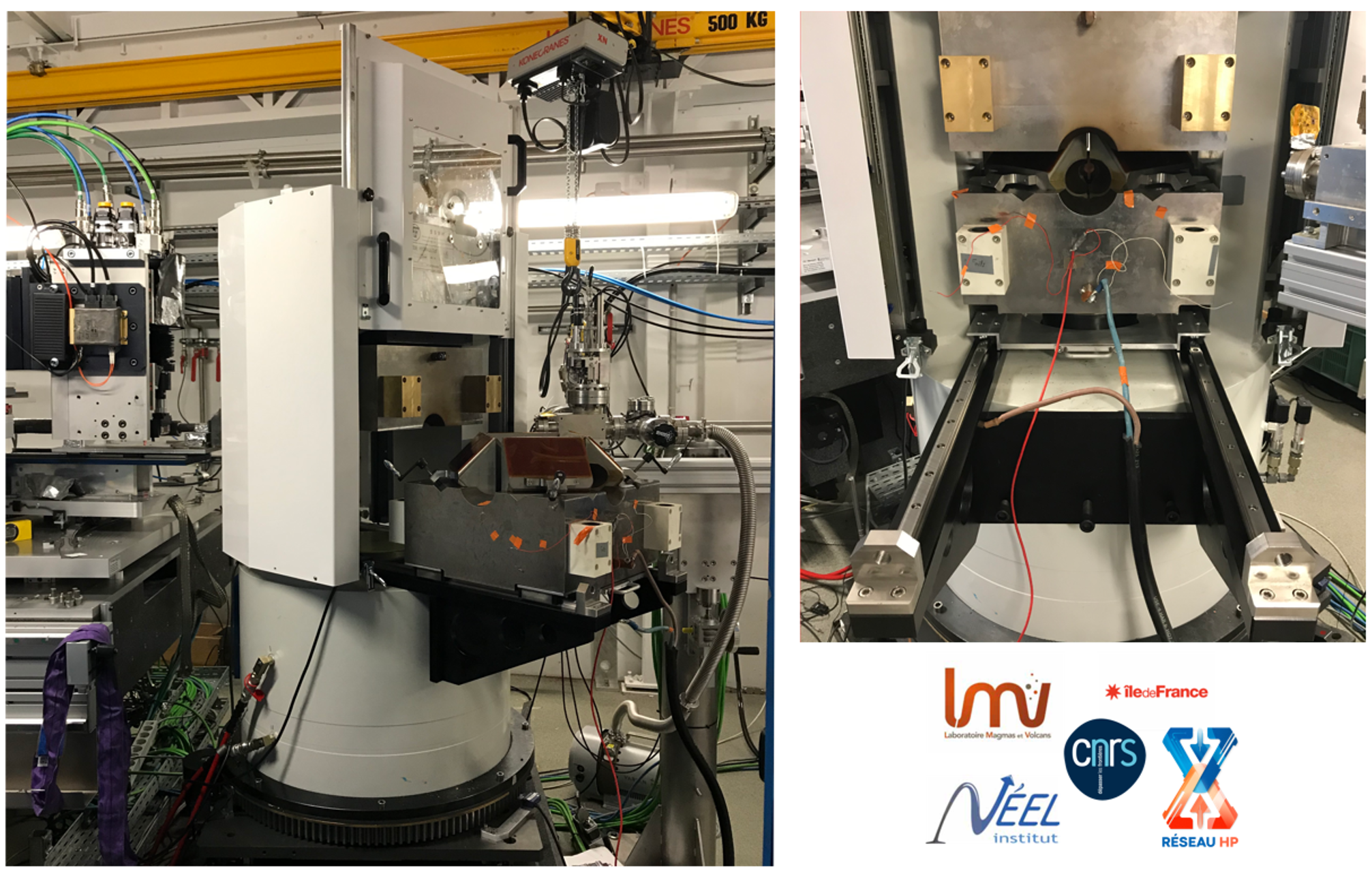
The multi-anvil press installed at PSICHÉ. (credits: Nicolas Guignot)
Contact: laura.henry@synchrotron-soleil.fr