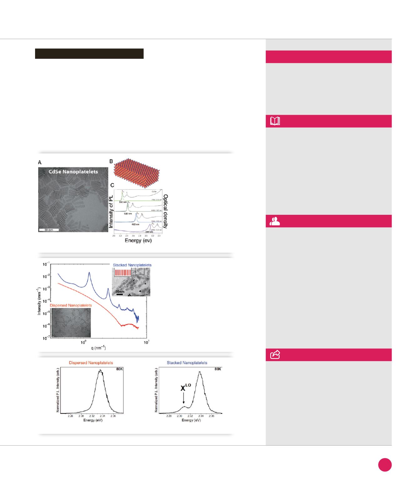
This stacking has important consequences
on the emission properties of the
nanoplatelets (figure
➌
). While a unique
peak is visible in the emission spectra
of pure hexane dispersion, a low energy
line emerges when an anti-solvent is
added. A similar line is also visible when
solutions are dried on a substrate. In
order to unravel the physical mechanism
at play for the emergence of this low-
energy peak, advanced spectroscopy
experiments have been performed. They
showed that this band could be attributed
to the longitudinal optical (LO) phonon
replica of the band-edge exciton. The
appearance of the band in self-assembled
nanoplatelets is explained using a model
based on an efficient photon reabsorption
between neighboring nanoplatelets. These
finding could have potential applications
in optoelectronics devices such as lasers
based on the confined mode of this
phonon replica.
Phonon line emission revealed
SWING beamline
ASSOCIATED PUBLICATION
Phonon Line Emission Revealed
by Self-Assembly of Colloidal Nanoplatelets
M.D. Tessier, L. Biadala, C. Bouet, S. Ithurria,
B. Abecassis, and B. Dubertret
ACS Nano 7(4) (2013) 3332
REFERENCES
[1] S. Ithurria & B. Dubertret, J. Am. Chem. Soc.
130 (2008), 16504
[2] S. Ithurria et al. Nat. Mater. 10 (2011), 936
Laboratoire de Physique des Solides, Bât 510,
Université Paris-Sud, 91405 Orsay, France
CORRESPONDING AUTHOR
➊
A
) TEM image
of CdSe nanoplatelets.
B
)
Schematic representation.
C
) Thickness dependent
optical properties of
nanoplatelets.
➋
SAXS patterns of
dispersed and stacked
nanoplatelets acquired
on the SWING beam line.
➌
Emission spectra at 80K of dispersed and stacked platelets in solution.
47
SYNCHROTRON
HIGHLIGHTS
2013


