Bone growth regulation:
how does osteopontin
bind uranium?
The impact of radioelements on living
organisms and human has been studied
since the nineteen fifties and their chemical
toxicity is thought to resemble heavy metal
toxicity by replacement of essential metals
of the organism such as iron or calcium.
In the case of uranium, one challenge to
better appraise its toxicity and develop
countermeasures in case of exposure
of living organisms is to better assess
pathways of contamination. Among others,
the skeleton is a targeted deposition site of
this element whatever the oxidation states
that have been administered to animals.
Radioelements in general are chemical
and radiological contaminants. It is known
that chemical toxicity prevails in the case
of uranium and thorium, while both
radio- and chemical toxicity are important
for plutonium. In the case
of chronic and very low level exposure,
the clinical consequences are still largely
undefined [1]. Whatever the exposure
pathway (inhalation, ingestion or wound),
the soluble fractions of radioelement are
absorbed and transported by blood and
are either evacuated through renal
excretion, or fixed in target organs. Liver
is the most important soft tissue deposition
site for all of them except uranium,
for which it is the kidneys [2].
On internal contamination, uranium enters
the blood stream in the form of soluble
uranyl (the dioxo cationic form U(VI)O
2
2+
)
salts and protein-bound complexes and
is then mainly deposited in kidney and
bone tissues. It was recently suggested
that chronic mechanisms of uranyl toxicity
might include phosphate interactions
which could explain its
in-vivo
targets [3].
Recently, Quémeneur
et al.
suggested
the use of osteopontin (OPN) as a possible
uranyl exposure biomarker observing
the reduced concentration of OPN in urine
upon exposure [4]. Screening several
peptides present in its sequence led
to the identification of a phosphorylated
hexapeptide pSDEpSDE as the most potent
calcium growth inhibitor and thus
as one possible OPN active site.
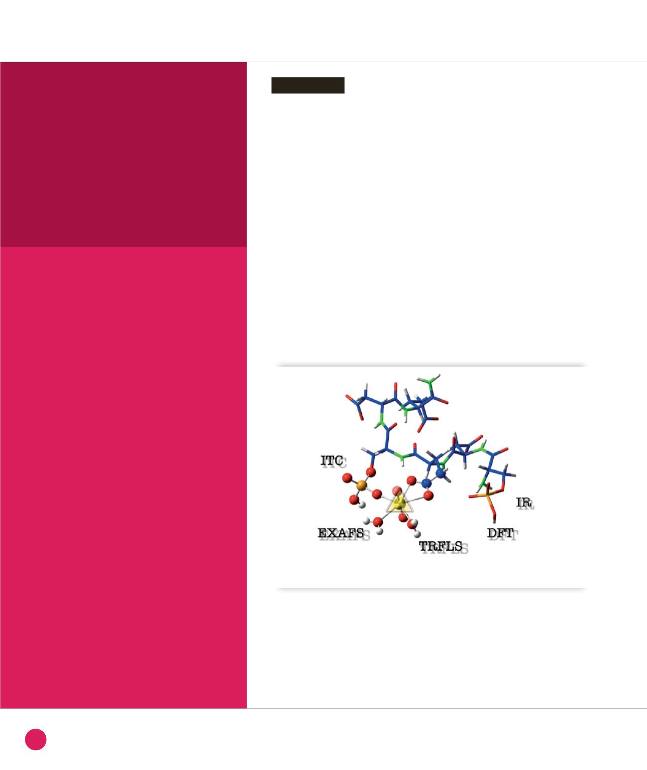
We have investigated the structural
aspects of the uranyl complexes
formed with the pSDEpSDE peptide
and the protein by a combination
of techniques (calorimetric titration,
vibrational spectroscopy, X-ray Absorption
Spectroscopy) and quantum chemical
calculations as schematized in Figure
➊
.
Introduction
CHEMISTRY AND PHYSICAL CHEMISTRY, NANOCHEMISTRY
➊
Molecular simulation model of the uranyl-pSDEpSDE complex.
40
SYNCHROTRON
HIGHLIGHTS
2013


