Our results show the structural
polymorphism of inclusions in HD brain.
We propose that the inclusions lacking
any structural rearrangement constitute
nontoxic amorphous aggregates, whereas
the amyloid inclusions enriched in both
β
-sheet and
β
-sheet/unordered are highly
neurotoxic, as they are always associated
with the most severe form of the disease
and found in the most affected brain
regions.
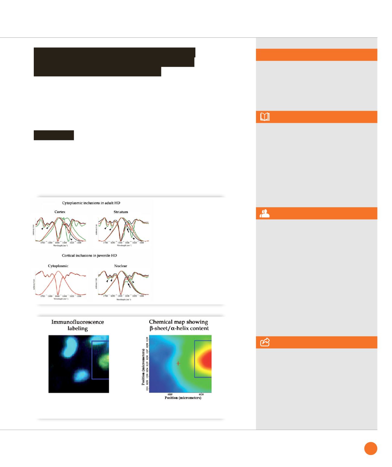
We collected spectra centered on Nis and
nuclei without inclusions (as controls) in
the cortex and striatum of three juvenile
HD cases. We demonstrated enrichment in
β
-sheets (1627, 1681 and 1693 cm
-1
) in this
category of inclusions (Fig.
➋
). Because
one of the main contributions was at
1627 cm
-1
, we conclude that juvenile Nis
are amyloid. We also observed that they
shared with adult Cis of striatum, but not
those of cortex, the
β
-sheet/unordered
enrichment in the component at 1639 cm
-1
.
Conclusion
Nuclear inclusions in juvenile HD cases possess
an amyloid structure resembling that of striatal
cytoplasmic inclusions in adult cases
SMIS beamline
ASSOCIATED PUBLICATION
Structure of inclusions of Huntington’s disease
brain revealed by synchrotron infrared
microspectroscopy : polymorphism
and relevance to cytotoxicity
W. André, C. Sandt, P. Dumas,
P. Djian and G. Hoffner*
Analytical Chemistry 85(7) (2013), 3765
REFERENCES
[1] Nekooki-Machida et al. Proc Natl Acad Sci
U S A 106 (2009), 9679
2] The Huntington’s Disease Collaborative
Research Group. Cell 72 (1993), 971
[3] J. Vonsattel et al. J Neuropathol Exp Neurol.
44 (1985), 559
[4] M. DiFiglia et al. Science. 277 (1997), 1990
[5] Nilsson, MR. Methods, 34 (2004), 151
* Laboratoire de Physiologie Cérébrale,
UMR 8118, Université Paris Descartes,
45 rue des Saints-Pères, 75006 Paris, France.
CORRESPONDING AUTHOR
➌
Comparison of the immunofluorescence labeling and the chemical mapping of an area containing an amyloid
inclusion, showing the correspondence between the inclusion (green, left panel) and the area with the highest
β
-sheet content (red, right panel).
➋
Comparison of spectra of cytoplasmic
or nuclear inclusions with spectra
of controls (inclusion-free cytoplasm
and nuclei) in adult and juvenile cases,
showing enrichments in
β
-sheets
(1627 cm
-1
, 1681 cm
-1
, and 1693 cm
-1
;
arrowheads) and enrichments in
β
-sheet/
unordered structure (1639 cm
-1
; arrow)
in some inclusions. The analysis reveals
the amyloid (1627 cm
-1
) or amorphous
nature of inclusions.
55
SYNCHROTRON
HIGHLIGHTS
2013


