Bienvenue à PROXIMA 2A !
Une lumière pour la biologie et la micro-cristallographie.
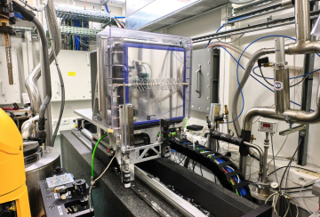
PROXIMA 2A (PX2-A) est une ligne de lumière micro-foyer-X dédiée à la bio-cristallographie et aux innovations méthodologiques pour les microfaisceaux-X. Ouvert depuis 2013, les sujets traités vont au delà de la cristallographie standard des protéines et incluent la découvert des médicaments, la résolution de protéines membranaires, la virologie, la cristallographie des petites molécules, la diffraction-X des poudres et même la cristallurie. La ligne de lumière est extrêmement automatisée et conçue pour aider les scientifiques à s’attaquer aux cibles structuraux et aux systèmes biologiques les plus exigeants. L’énergie des rayons-X est facilement accordable sur une grande gamme (6 – 18 keV) et rend la plupart des seuils d’absorption accessibles pour des expériences en diffraction-X anomale. La station expérimentale est équipée d’un passeur d’échantillon de haute capacité (148 ports d’échantillon du type SPINE), un goniomètre-kappa de haute performance, un détecteur de fluorescence-X (KETEK) et un grand détecteur-bidimensionnel rapide de faible bruit et compteur de photons unique - l’EIGER X 9M (dont 238 images par second en mode 9M ou 750 images par second en mode 4M).
See also Biology Health : HelioBio
L'équipe
Nos derniers tweets
Offres d'emploi
Contacter William SHEPARD pour plus d'information.
Posters
- "From Plate Screening to Artificial Intelligence: Innovative developments on PROXIMA 2A at Synchrotron SOLEIL" - Mechanical Engineering Design of Synchrotron Radiation Equipment and Instrumentation (MEDSI 2018), 25-29 June 2018. Paris, France.
- "The CRIBLEUR: An Any Format Screener for in situ X-ray Diffraction Experiments on PROXIMA 2A" - Biologie Structurale Integrative (BSI), 29 Nov - 03 Dec 2021, Paris-Saclay, France.
Présentations
- Of Symmetry, Lattices & Space Groups - ReNaFoBis Webinar Series, 09 Dec 2020, Paris, France
- PROXIMA 2A Tutorial: Best Practices in the use of Micro-beams in Macromolecular Crystallography - SOLEIL Users Meeting (SUM2023), 20 Jan 2023, Synchrotron SOLEIL, Gif-sur-Yvette, France
- Living Machines @ Work : As seen by Macromolecular Cristallography - LM@W day at I2BC, CNRS, Gif-sur-Yvette, France
- Some Theoretical Aspects of Serial Crystallography - RECI-TECH 2025, 8-11 Dec 2025, Synchrotron SOLIEL, Saint Aubin, France
Vidéos
- Introducing PROXIMA-2A (http://tinyurl.com/PROXIMA-2A-VIDEO)
- In-situ crystal screening and data collection with the "Cribleur" (http://tinyurl.com/CRIBLEUR-VIDEO)
Commenter Citer PROXIMA 2A
Les publications de la ligne de lumière PROXIMA 2A à citer sont listées ci-dessous:
- The general PROXIMA 2A beamline publication is in the SRI 2013 Proceedings.
- Duran et al. (2013) "PROXIMA 2A – A New Fully Tunable Micro-focus Beamline for Macromolecular Crystallography" Journal of Physics: Conference Series 425, 012005. doi:10.1088/1742-6596/425/1/012005.
- Martin Savko's oral presentation on using EIGER detectors at the ECM 30 (Basel, Switzerland).
- Savko et al. (2016) "Getting the most of the sample with small beams and fast detectors." Acta Cryst A, a54283. https://doi.org/10.1107/S2053273316097163
- Studies on using Helical Scans to mitigate X-ray radiation damage is published in a special issue of the Journal of Synchrotron Radiation.
- Polsinelli et al. (2017) "Comparison of helical scan and standard rotation methods in single-crystal X-ray data collection strategies." J. Synchrotron Rad. 24, 42–52. https://doi.org/10.1107/S1600577516018488.
- Adam Simpkin's SIMBAD pipeline is described in the Act Cryst D publication below.
- Simpkin et al. (2018) "SIMBAD: a sequence-independent molecular-replacement pipeline." Acta Crystallogr D Struct Biol. 74, 595–605. https://doi.org/10.1107/S2059798318005752.
- The Cribleur plate screener is described in the MEDSI 2018 Proceedings.
- Jeangerard et al (2018) "FROM PLATE SCREENING TO ARTIFICIAL INTELLIGENCE: INNOVATIVE DEVELOPMENTS ON PROXIMA 2A AT SYNCHROTRON SOLEIL." JACoW Publishing, MEDSI 2018 Proceedings WEPH36. https://doi.org/10.18429/JACoW-MEDSI2018-WEPH36.
- Studies on fast data collections with the Eiger X 1M for the ELI beamlines (Prague, Czech Republic).
- Khakurel et al. (2020) "Kilohertz Macromolecular Crystallography Using an EIGER Detector at Low X-ray Fluxes." Crystals 10, 1146. https://doi.org/10.3390/cryst10121146.
- Developing Synchrotron Serial Crystallography methods for MOFs, POMs and small unit cell crystals.
- de Zitter et al. (2024) "Elucidating metal–organic framework structures using synchrotron serial crystallography." CrystEngComm, advance publication. https://doi.org/10.1039/d4ce00735b.
Données techniques
6 - 18 keV
~0.0002 (Si 111)
Onduleur "canted" U24 (4.5 mrad)
Faisceau blanc/rose
Monochromateur "channel-cut" refroidit par l'azôte liquide, (miroir convxe prefocalisant), miroirs focalisants en configuration Kirkpatrick-Baez.
Linéaire
EIGER X 9M area detector for X-ray diffraction images (238 fps in 9M mode, 750 fps in 4M mode)
Ketek SDD detector for X-ray fluorescence scans
Diamond scCVD and Ti foil XBPMs
High resolution X-ray camera (SOLEIL in-house design)
Thématiques scientifiques
| Micro-crystallography |
Small and weakly diffracting crystals |
|---|---|
| SAD and MAD phasing |
Phasing with a range of heavy atoms, including sulphur |
| Plate screening |
Screening of crystallization trays |
En recherche interne à SOLEIL, la ligne de lumière PROXIMA-2A est rattachée à la section scientifique :
| Section scientifique SOLEIL | Biologie, santé, Héliobio |
|---|
Mise à jours
2023-05-26 Nouveau server pour l'acces à distance, nouvelle station de travail MXCuBE et nouvelle documentation
- Il y a un nnouveau server pour l'acces à distance: remote.synchrotron-soleil.fr:4000, protocol NX.
- Il y a aussi une nouvelle station de travail MXCuBE: proxima2a-pc4
- Voici la documentation mise à jour:
2023-01-20 Presentation du Tutoriel de PROXIMA 2A
- PROXIMA 2A Tutorial: Best Practices in the use of Micro-beams in Macromolecular Crystallography - SOLEIL Users Meeting (SUM2023), 20 Jan 2023, Synchrotron SOLEIL, Gif-sur-Yvette, France
2022-10-11 HDF5 structure & fix_transformation_negative.py
- Un bug avec le fichier master HDF5 de l'EIGER X 9M est lié à un valeur négatif dans la transformation du detecteur - specifiquement /entry/instrument/detector/transformations/translation[2]
- Le command sur la machine process1 pour corriger la valeur negatif est:
- fix_transformation_negative.py my-project_master.h5
- Notez ce command modifies directement le parametre dans le fichier master HDF5, il ne crée pas un nouveau copie. Si la parametre est deja positif, il reste positif.
- XIA2 et DIALS doivent fonctioner à nouveau maintainant
2022-06-13 ADXV & ALBULA
- ADXV et ADXV_FOLLOW sont disponibles pour visualiser les images de diffraction. Dans ADXV, il faut d'abord cliquer sur le fichier MASTER pour telecharger les parametres de la collecte (distance, longueur d'onde, centre du faisceau, etc...). Les commandes sont :
- adxv
- adxv_follow
- Avec les modifications de la format HDF5, ALBULA n'affiche pas tous les images de diffraction sur la sation de travail proxima2a-10. Pendant qu'on resoudre la probleme, merci d'utiliser ADXV et ADXV_FOLLOW.
2022-06-13 Generation automatique des fichiers CBF
- Des fichiers CBF sont automatiquement generés et stockés dans le sous-reproratoire ${prefix}_cbf.
2022-06-02 EIGER Firmware Update
- Suite à une mise à jour du logiciel interne de l'EIGER 9M, la structure des fichiers HDF5 a changé un peu, et le module d'extension Neggia (version 0.6) qui traites les anciens et nouveaux fichier HDF5 peut se trouver au lien suivant: https://bit.ly/dectris_neggia_0p6_linux. Contactez-nous si vous avez des problemes pour traiter les données.
- autoPROC - il devrait marcher si on utilise le bon module d'extension Neggia
-
xdsme - il faut modifer deux lignes dans XIO.py:
-
"import pyfive" -> "import h5py"
-
self.rawHead = pyfive.File(self.fileName) -> self.rawHead = h5py.File(self.fileName, 'r')
-
Calendier PX2-A
Cliquer sur le lien ci-dessous pour voir les dates du temps de faisceau-X disponible sur PX2-A:
- AP36 (septembre 2025 - mars 2026) cliquer ici, ou http://tinyurl.com/PROXIMA-SOLEIL-AP36
Tutoriels & Guides
Tutoriels
Lisez le tutoriel de PROXIMA 2A (SUM 20-21 janvier 2023):
Guides
Voir aussi les sections Posters & Presentations et DOWNLOADS à la fin de la page.
Avant de visiter PX2-A
Merci de contacter votre "Local Contact" avant votre session.
- Combien des échantillons?
- Prevoir 2 heures par Uni-puck (16 SPINE pins)
- 1 shift = 8 heures = 4 Uni-pucks
- 2 shifts = 16 heures = 8 Uni-pucks
- Prevoir 2 heures par Uni-puck (16 SPINE pins)
- Quels longueur d'ondes, energies ou derivés d'atomes lourdes?
- Energie nominale de la ligne = 12.650 keV
- Gamme d'energie : 6 - 18 keV
Dewars
Il y a DEUX moyens pour transporter les Dewars vers SOLEIL:
- déposer/enlever à la main au Magasin de SOLEIL
- pour les labos en Ile-de-France
- livrer/enlever par un transporteur avec BORDEREAUX PRÉ-PAYÉS (DHL, FEDEX, UPS, etc...)
- pour les labos français hors Ile-de-France
- pour les labos hors France
Les transports des Dewars pouront etre prepayés par SOLEIL ou iNEXT Discovery (SOLEIL vous enverra les bordereaux prépayés):
- pour les labos français hors Ile-de-France par SOLEIL jusqu'au trois (3) Dewars par session (que la session soit 1, 2 ou 3 shifts)
- Contacter le User Office de SOLEIL (UserOffice@synchrotron-soleil.fr)
- numero de projet SOLEIL (e.g. 20191234)
- numero de la session (e.g. #3)
- Contacter le User Office de SOLEIL (UserOffice@synchrotron-soleil.fr)
- pour les labos hors France par iNEXT Discovery jusqu'un (1) Dewar par session (que la session soit 1, 2 ou 3 shifts)
- Contacter le User Office de SOLEIL (UserOffice@synchrotron-soleil.fr)
- numero de projet SOLEIL (e.g. 20191234)
- numero PID d'iNEXT (e.g. PID 13476)
- numero de la session (e.g. #3)
- Contacter le User Office de SOLEIL (UserOffice@synchrotron-soleil.fr)
Le contenu de Dewar:
- UN BORDEREAUX PRÉ-PAYÉ POUR LE RETOUR.
- une disque dur externe (HDD 1-2 To) pour le REMOTE ACCESS
Envoyez les Dewars aux Local Contacts (l'un des scientifiques ci-dessous):
- Willima SHEPARD (Responsable ligne de lumière)
- Serena SIRIGU
- Martin SAVKO
Les numéros de téléphones de la ligne de lumière PROXIMA 2A (pour le transport des Dewars):
- +33-1-6935-8180 (fixe)
- +33-1-6935-8181 (sans-fils)
L'addresse de SOLEIL:
PROXIMA 2A
Synchrotron SOLEIL
L'Orme des Merisiers
Départementale 128
91190 SAINT-AUBIN
FRANCE
Merci de prevenir votre Local Contact, puisqu'il(elle) pourrait remplir votre Dewar avec de l'azôte liquide quand il arrive!
Accès à Distance (Remote)
Acces à Distance (Remote)
Attention! Le server NoMachine et la station de travail MXCuBE ont changé depuis avril 2023:
- En bref pour connecter via NoMachine
- Créer ou ouvrir une connection NoMachine
- SERVER = remote.synchrotron-soleil.fr
- PORT = 4000
- PROTOCOL = NX
- Se logger avec votre identifiant du projet et mot-de-passe
- Connecter à la nouvelle station de travail: proxima2a-pc4
- Ouvrir une session "Custom Session" (non pas "Physical Display")
- Quand la fenetre terminal-X apparaisse, vous pourriez changer le "keyboard map" du clavier. Tapper:
- setxkbmap fr [si votre clavier est AZERTY]
- setxkbmap us [si votre clavier est QWERTY]
- Tapper CNTL-SHIFT-T pour ouvrir un nouveau onglet / terminal-X
- Tapper dans quatre onglets distinct
- mxcube
- firefox -P
- albula ou adxv_follow
- ssh -X process1
- Créer ou ouvrir une connection NoMachine
Sinon, merci de lire la documentation disponible (en anglais) par le lien :
- https://drive.google.com/file/d/1cKLEgct9CGid8n5vA2bDEsgAVQq2eXte/view?usp=share_link
- L'ancien version est ici:
- https://drive.google.com/file/d/1J-Ez8_sGF3k28khLhSgAEF9Mrs7pKVy7/view?usp=sharing
- L'ancien version est ici:
Verifier que vous avez les equipement suivants:
- une version recent de NoMachine
- une bonne connection internet (ADSL ou fibre optique)
- Un clavier QWERTY
- sinon utiliser le command "setxkbmap fr"
- un souris à trois (3) boutons pour le centrage des cristaux
- un écran haute resolution (2560 * 1440 pixels)
Pour tester la connection avant votre session sur PROXIMA 2A
- Contacter l'equipe de la ligne pour organiser une session test
Pour connecter à distance:
- Connectez-vous via NoMachine
- HOST: remote.synchrotron-soleil.fr
- PORT = 4000
- PROTOCOL = NX
- Se logger sur remote.synchrotron-soleil.fr avec votre numero de projet et son mot-de-passe, par exemple:
- User name = 20230123
- Password = 43jR89pL67QX
- Choisir le station de travail :
- proxima2a-pc4
- Connecter via "Custom Session "
- Attendez qu'un gnome terminal apparaisse :
- Creer quatre onlets, <shift-cntl-t>
- Lancer les commandes dans les onglets
- firefox -P
- mxcube
- adxv_follow ou albula
- ssh -X process1
==========================
DewarsLabel your Dewar properly
Your name, address & email
Project Number
Beamline
Local Contact or BL Manager
Include the following with your Dewar
a Hard Disk Drive (HDD 1-2 TB)
a Return Shipping Label or Transporter Account Number
Send or drop off your Dewar, which must arrive:
either 24 hours before your session
or the Friday morning for weekend sessions
Dewars may be dropped off at the SOLEIL gate
Types des echantillons
PROXIMA-2A prends tous les echantillons - les grands comme les petits! Et même les cristaux pas très beaux...
- grand > 10 microns
- petit < 10 microns
- pas beau = fissuré, fracturé ou agglutinés, car souvent se sont des amas des micro-cristaux!
Types des port d'echantillons :
Robot CATS - Uniquement des pins du type SPINE dans des Uni-pucks
- l'echangeur du CATS peuvent contenir jusqu'au neuf (9) uni-pucks, et chaque uni-puck peut contenir 16 SPINE pins, dont un total de 144 pins pour le grand Dewar du CATS sur PX2-A
- Les pins SPINE sont disponibles chez Hampton, Mitegen & Molecular Dimensions
- longueur du pin = 22 mm (du boucle au base)
- deplacement horizontale possible du goniometre = -25 to +5 mm
Montage manuel
- Le "mini-kappa" du goniometre peut etre incliné pour permettre un transfert facile sous cryogenie:
- Mettez omega = 0° and kappa = 30 - 60°
- Un video expliquant le transfert des echantillons dans les fioles (vials) vers les Uni-pucks.
- Un video demontrant comment utiliser un "dry shipper" et comment recuperer les echantillons :
Pendant vos experiences
- Debut & fin pour les seesions sur PROXIMA-2A :
- trois (3) shifts = 8h30 – 8h00 (le lendemain)
- deux (2) shifts = 16h30 - 8h00 (le lendemain)
- un (1) shift = 8h30 - 16h00
- L'equipement & accessories disponible aux utilisateurs:
- Microscopes, pins SPINE, boucles & wands for fishing crystals
- Materiel pour la cryogenie - azôte liquide, Dewars pour manupler les uni-pucks, cryo-tongs, gants, etc...
- TOUS LES EQUIPEMENT DOIVENT RESTER SUR PROXIMA 2A!
- Les acquisitions des données sont faites via MXCuBE , voir le lien ci-dessous pour le guide (PX2-A MXCuBE Users Guide):
- Les fonctionalités
- Verification de la position du faisceau-X (BeamCheck)
- Transfert des echantillons (MOUNT & UNMOUNT)
- Centrage des cristaux
- Collectes des données cristallographiques
- Changement de l'energie du faisceau-X
- Spectra XFE et balayage d'energie
- Collectes SAD & MAD
- Collectes helicoidales
- Balayages GRID et MESH
- Energies du faisceau-X disponibles
- 6 – 18 keV
- Seuils d'absorption disponibles
- K-edges Cr – Zr
- L3-edges Ce - U
- Temps typiques des collectes
- Minimum exposure time 4.2017 ms per frame (238 fps), nominal exposure time 4.5 ms per frame (222 fps)
- 16.2 s per nominal data collection rate (4.5 ms par image, 0.1 degrees par image, 3600 images)
- Attention: les 16.2 s n'inclus pas le mis en place du beamstop, movement du detecteur, ouverture de l'obturateur, etc...
- Les parametres de default sont bien pour le plupart des cristaux
- Pour visualiser les images de diffraction-X
- ALBULA
- ADXV
- Traitement des images de diffraction-X
- XDSME
- autoPROC
- XIA2
- Horaires d'ouverture de la cantine
- Lundi - Vendredi :
- Petit dejeuner = 7:30 - 9:00 ,
- Dejeuner = 11:45 - 13:45
- Dîner = 19:00 - 21:00 (fermature 21:30)
- Weekends (Samedis et Dimanches) & Jours de fetes :
- Petit dejeuner = 7:30 - 9:00
- Dejeuner = 12:30 - 13:30
- Dîner = 19:00 - 21:00 (fermature 21:30)
A la fin de vos experiences
- Repatriation des données
- Telecharger vos données sur une disque dur externe via un port USB3 ou USB2
- Si possible, utiliser le script automatisé "altsync" (cntrl-C pour l'arreter et puis demounter le disque externe).
- Uniquement les petites volumes des données (e.g. fichiers MTZ) peuvent etre telecharger chez vous via le systeme" SOLEIL Data Retrieval"
- Enlever l'echantillon du goniometre, enveler tous les uni-pucks et fermer les couvercles du CATS
- Signer le "Safety Approval Sheet"
Important:
- Merci de nous dire vos reactions et remarques pour améliorer PROXIMA 2A!
- Merci de nous envoyer des images, diaposatifs, publications de vos resultats!
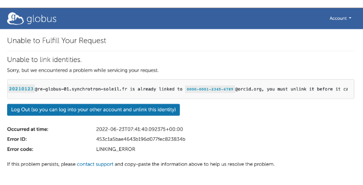
Recuperation des données via GLOBUS
Vous pouvez telecharger vos données via GLOBUS. Voici quelques conseils:
- consulter la documentation GLOBUS (mis à jour le 22 juin 2023)
- utiliser une connection internet assez rapide
- e.g. Free Box avec fibre optique
- compter 40 minutes pour 100 GB
- e.g. Free Box avec fibre optique
- arreter le VPN qui bloque le telechargement du cle de securité GLOBUS
- e.g. SecurePulse
- utiliser votre compte ORCID
- e.g. ORCID #0000-0001-2345-6789, si vous ne le avez pas, créer un.
- il faut le user_id de votre compte projet SOLEIL
- e.g. 20220123 [we02LK98]
- Desactiver les Screen Saver et le Power Nap
- Ils peuvent interrompre le telechargement
- IMPORTANT: Après le transfert des données, merci de rappeler de deconnecter votre lien (data link)!!!
- "Unable to link identities" .... "20220123@re-globus-01.synchrotron-soleil.fr is already linked to 0000-0001-2345-6789@orcid.org, you must unlink it..."
- Voir la fin de la Section 7 de la documentation de Globus.