Scientists reveal the third dimension of magnetic skyrmion tubes
An international team of researchers from the Max Planck Institute for Intelligent Systems in Stuttgart, the Universities of Southampton, Exeter, Warwick and Cambridge, and 3 synchrotrons -BESSY II, SOLEIL and Diamond- coordinated by Prof. Peter Hatton, Durham University, have succeeded in imaging the three dimensional structure of a skyrmion tube. This achievement will allow the nanoscale mechanisms governing the formation and destruction of skyrmions to be investigated. Their results are published in Nature Communications.
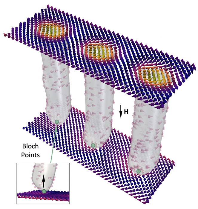
Skyrmions - topological, nanoscopic magnetic whirls - are commonly presented as two dimensional, disc-like objects. However in reality, they are believed to extend through the thickness of the material as an elongated string-like tube, as visualised in Figure 1. Due to the limitations of conventional magnetic imaging techniques, real-space imaging of this vertical dimension of the skyrmion spin texture has remained elusive.
Now, an international team of researchers from the UK, France and Germany have overcome these limitations using magnetic x-ray holography and microscopy techniques. “Previously, these skyrmion tubes had only been shown in simulation work,” explains Max Birch, a PhD student at Durham University, and the lead author on the publication. “We found a way to experimentally image this previously unexplored dimension of the skyrmion state.”
The topological properties of magnetic skyrmions mean that they are stable against all but the largest of deformations, giving them a potential use as information storage element in future data devices. However, efficient control of the reading and writing of skyrmions requires understanding of their formation and destruction mechanisms. The tube-like structure is the key starting point: skyrmions unwind by the motion of magnetic singularities, known as Bloch points, at the end of each skyrmion tube, acting as a topological zipper. Successful imaging of skyrmion tubes has now opened the door to further research into the dynamics governing these processes.
The researchers used 120 nanometer thin iron-germanium (FeGe) lamellae, prepared by focused ion beam milling, in which they were able to visualise the 3D structure of the skyrmion tubes. FeGe is one of a number of chiral magnets that have recently been discovered to host magnetic skyrmions.
Among other instruments, the COMET endstation at SEXTANTS beamline, was crucial in performing x-ray holography imaging techniques. “Holography allows us to recapture the phase information of a diffracted x-ray beam. Typically, this phase information is lost, meaning reconstructing an image of the sample from x-ray diffraction data is impossible,” explains Birch. “The SEXTANTS beamline at SOLEIL, with its ability to cool the sample down to cryogenic temperatures, is uniquely capable of performing these measurements.” Using this technique, the researchers were able to image skyrmions with only a 70 nm size, 100 times smaller than a human hair.
Birch’s supervisor, Prof. Peter Hatton, Durham University, brought together an international research team, involving scientists from the Max Planck Institute for Intelligent Systems in Stuttgart, the Universities of Southampton, Exeter, Warwick and Cambridge, and experts at the synchrotron radiation sources BESSY II in Germany, SOLEIL in France and the Diamond Light Source in Great Britain. “The project truly was an interdisciplinary team effort, with contributions from a wide range of experts in crystal growth, micromagnetic simulations, and x-ray imaging methods,” says Hatton. Birch concludes, "The ability to bring together multiple X-ray instruments and techniques, and take advantage of their unique capabilities, was crucial to the success of this project".