Revealing the 3D chemical composition of whole fossils
An international team led by the IPANEMA laboratory (CNRS / French ministry of Culture / Univ Versailles St-Quentin) and GALAXIES beamline of SOLEIL publishes a new technique for studying the composition of entire fossils in the journal Science Advances. Synchrotron 3D X-ray Raman imaging provides the composition of entire fossils at a scale of one hundredth of a millimeter. Applied to a 53-million-year-old ant preserved in amber, the team reveals the presence of chitin that constituted the insect's exoskeleton and even visualizes a difference in preservation between the part of the insect that was first in contact with the resin and the part that was only later.
An international team led by the IPANEMA laboratory (Paris-Saclay), and the Synchrotron SOLEIL, in collaboration with three other CNRS laboratories (ISYEB, IMPMC and IRCP), the University of Lausanne and two synchrotron facilities (ESRF and SLAC) has just shown that it is possible to establish the composition at each point of an organic fossil based on the totally innovative use of a method resulting from fundamental research in X-ray spectroscopy.
The study of fossil materials has benefited from 3D X-ray computed tomography for about fifteen years, comparable to medical scanning. Tomography allows the study of microscopic patterns necessary to understand the evolution of species, their physiology and fossilization mechanisms. It leads to "black and white" images indicative of the density of materials, as a medical X-ray shows the relative absorption of different tissues, but does not provide the internal molecular composition of fossils. Other methods, such as X-ray fluorescence imaging can produce an image of the chemical composition, but only on flat fossils.
With 3D X-ray Raman imaging, a small fraction of the energy of the X-rays used to illuminate a material is absorbed by its constituting atoms. These will emit a signal, specific of each type of atom, permitting to determine the chemical composition of the material. The team showed that coupling this method with advanced instrumentation using the qualities of focusing, extreme accuracy in the chosen wavelength, and penetration of synchrotron X-rays allowed the chemical composition to be obtained at each point of the fossil, in 2D or 3D.
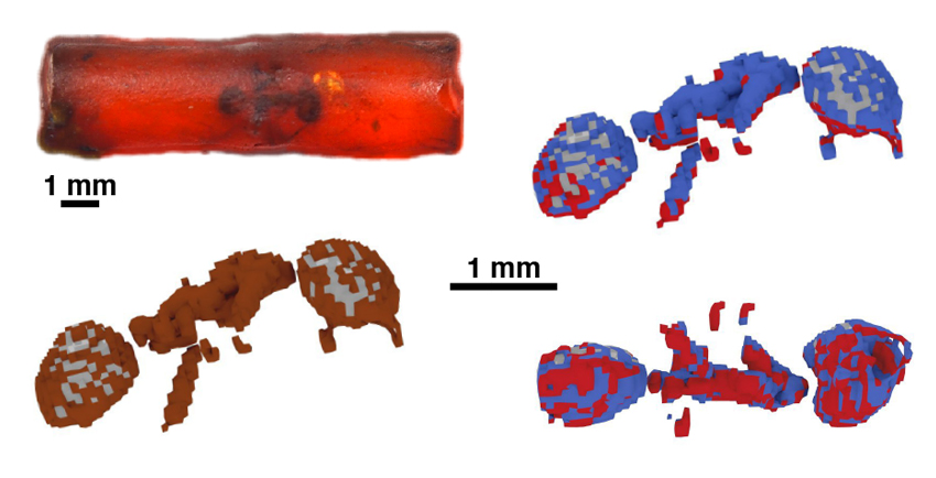
The team showed that a 53-million-year-old fossil ant exceptionally preserved in amber had retained traces of chitin, an extremely resistant complex sugar used by insects as the main structural component of their exoskeleton. The researchers were able to show that the surface of the insect that had been brought into contact first with resin had been chemically preserved better than the one that had been covered later after the insect's death. These results are published in the journal Science Advances.