Investigation on the bioresorbable coronary scaffold resorption in-vivo
Coronary heart disease (CHD), the main cause for sudden deaths, is today treated with expandable stents to open the arteries for blood flow. But these stents cannot be surgically removed, and patients need medication to prevent blood clots. Magnesium scaffolds, however, are completely resorbed in the body, leaving only a remodeled artery that functions without a permanent cage. On the ANATOMIX beamline at SOLEIL, X-ray microtomography (µCT) imaging has shed light on the resorption process and biological response to the scaffold.
Bioresorbable magnesium stents (scaffolds) are a promising future treatment option for coronary artery stenosis, especially for young adults. Due to the resorption of these scaffolds (< 1 year), long-term interventions could be reduced compared to treatments with conventional, permanently lasting drug eluting stents1. First clinical trials investigating bioresorbable scaffolds indicate a return of vasomotion (i.e., the natural oscillations in contraction of the muscles in the blood vessel walls) after one year, which may be associated with improved long-term clinical outcomes. However, even after decades of development, the resorption process, ideal resorption time, and biological response in vivo are still not fully understood.
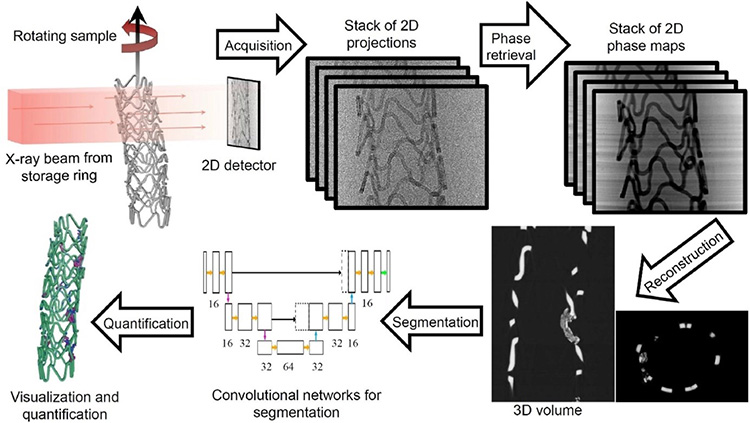
A recent study on the ANATOMIX beamline investigated the degradation of bioresorbable magnesium scaffolds in the coronary arteries of pigs, focusing on the influence of the thickness of the struts (i.e., the small segments forming the scaffold), and the presence of antiproliferative drugs. Due to the high sensitivity of synchrotron radiation microtomography with phase contrast (SR-µCT), the resorption of magnesium scaffolds could be evaluated qualitatively and quantitatively. To segment the µCT images a convolutional network architecture (U-net) was used. Due to the complexity of the 3D corrosion morphology, the analysis of large sample volumes would not have been possible without this deep-learning approach, or it would have required unrealistic amounts of human work.
In total, 30 bioresorbable magnesium scaffolds, made of the rare earth alloy Resoloy®, with different strut designs were implanted into the coronary arteries of 10 domestic pigs for 28 days using drug-coated or uncoated angioplasty balloons for post-dilatation. The resorption was analyzed using scanning electron microscopy, energy dispersive X-ray spectroscopy, and SR-µCT. The data from these methods were then related to the medical outcomes determined by angiography, optical coherence tomography and histology.
Besides giving new insights into the impact of strut design and drug coating on the resorption rate, this study demonstrates the tremendous potential of merging high resolution SR-μCT with deep learning methods for data analysis.
Based on the findings of the study, a new set of bioresorbable magnesium scaffolds has been designed and is used to further prepare the medical device certification process. The new scaffolds are explanted after different implantation lengths of time, and the corrosion kinetics will be investigated by exploiting the workflow established in the presented study.
Once the optimal scaffold design has been identified, optimizing for the right kinetics of resorption versus tissue ingrowth, the scaffolds will become available for the first in-human studies, paving the way for routine clinical application and improving the quality of life for patients requiring treatment with coronary stents.
The ANATOMIX beamline offers high coherence and photon flux and allows for short scanning times to obtain a full 3D volume image with high definition – in the present study, a few minutes per scan. Together with a newly available sample changing device, and the established deep-learning based data analysis pipeline, this high-throughput approach can become a blueprint for pre-clinical studies requiring the analysis of many complex samples, thus improving and accelerating medical implant development.
Video showing the rendering of a scaffold in 3D. (c) XPLORAYTION GmbH & MeKo
-----------------------
1 - drug-eluting stent: a drug substance preventing arterial cell proliferation coats the stent, significantly reducing the risk of stenosis recurrence. This substance is embedded in a polymer, which is plated onto the stent's metal mesh.