Imaging complex magnetic patterns in 3D
Ferromagnetic materials* have a non-zero spontaneous magnetisation at the nanoscopic scale. However, due to the competition of forces at play in the material, the local magnetic moments can form complex patterns at the microscale. Observing these patterns in 2D is possible with a variety of laboratory instruments, but the 3rd dimension has remained elusive until very recently. A recent study, carried at the SEXTANTS beamline, has allowed the development of a new tomographic technique to observe magnetic patterns in 3D with 80 nm resolution.
The research field of magnetism has seen the emergence of an exciting topic in the last years: 3D nanomagnetism. While two-dimensional magnetism has been vastly studied, thanks to a large panel of characterisation techniques, the interest in 3D nanostructures is very recent. The additional dimension offers a lot of new ways to stack magnetic moments together, providing a new playground to look for novel properties. For instance, chirality, this property that differentiate your left hand from your right hand, does not exist as such in 2D!
However, “seeing is believing” and studying magnetism in 3D requires a suitable microscope, or more exactly a tomographic microscope capable of observing magnetisation patterns in the bulk of the sample. This is precisely what an international team has developed on the beamline SEXTANTS.
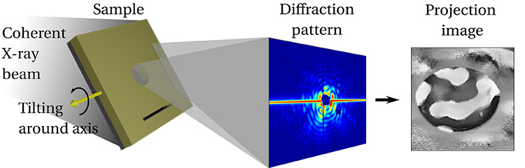
Their new magnetic tomography is based on a well-established microscopy method known as “Fourier-transform holography”. The idea behind holography is to create an interference between a reference wave and a wave traversing the object to be imaged. It is a bit as if you would place your object of interest just behind one of the slits in the famous Young’s double-slits experiment. The interference allows forming an image of the sample with a simple algorithm consisting essentially in a Fourier transform. To add magnetic contrast to this microscope, you exploit the unique properties of wavelength and polarisation of synchrotron radiation to capture the projection of the magnetic moments along the beam axis.
But all this gives you only 2D magnetic images! How can you do a Young’s double-slits experiment in 3D? Here is the trick implemented at beamline SEXTANTS: you exploit a natural tomographic axis perpendicular to the slit length and record projections of your sample at various angles, using the tilted slit as a reference for holography. But wait, magnetisation is a vector! If you rotate your vector around a single rotation axis, you are missing the component of the vector parallel to the rotation axis! Here is the second trick: add a second slit perpendicular to the first one, and record a second set of projections while rotating the sample around the length of the second slit.
With these two sets of projections and a good algorithm also developed by the team, you can compute the magnetic texture of your sample in 3D.
To demonstrate the effectiveness of the method, the team investigated a 1-µm thick multilayer made of iron and gadolinium.
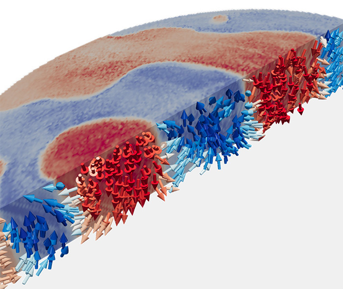
Laboratory microscopes showed a magnetic texture in curvy stripes with local magnetisation mostly up or down. The tomographic method allowed capturing the fine details of this pattern, in particular how the magnetisation rotates from up to down between neighbouring stripes. With 80 nm resolution to explore a 5 µm x 5 µm x 1 µm region, a lot of details are revealed!
*Ferromagnetic materials have the property of becoming magnetic themselves if placed in a magnetic field and keeping some of this magnetism when the field is removed.