Historical paintings at the nanoscale: L’Arlésienne (portrait of Madame Ginoux) by Van Gogh
A scientific collaboration between University of Antwerp (Belgium), the Kröller-Müller Museum (Netherlands) and the SMIS beamline of Synchrotron SOLEIL has presented the first nanoscale analysis of organic materials in historical paintings using optical‐photothermal infrared (OPTIR) microscopy. Focused on the Van Gogh’s painting L’Arlésienne (portrait of Madame Ginoux), this interdisciplinary team obtained striking results including the identification of the small red pigment particles used in the painting’s background as well as the characterization and distribution of the materials within the painting layers.
The chemical composition of cultural heritage objects is extremely heterogeneous due to the broad mixture of materials used for their elaboration, which also tend to react and transform with time producing additional substances. The identification of these chemical compounds, some of which are present in very low amounts, is key to understand the original materials and techniques used to produce the objects, as well as their history and conservation state which is an essential requirement to optimize the restoration and conservation strategies. Previous studies have shown the potential of high-resolution techniques such as X-ray based methods to characterize inorganic nanoparticles and layers. Nonetheless, there are still open questions regarding the most suitable method for the characterization of the organic layers.
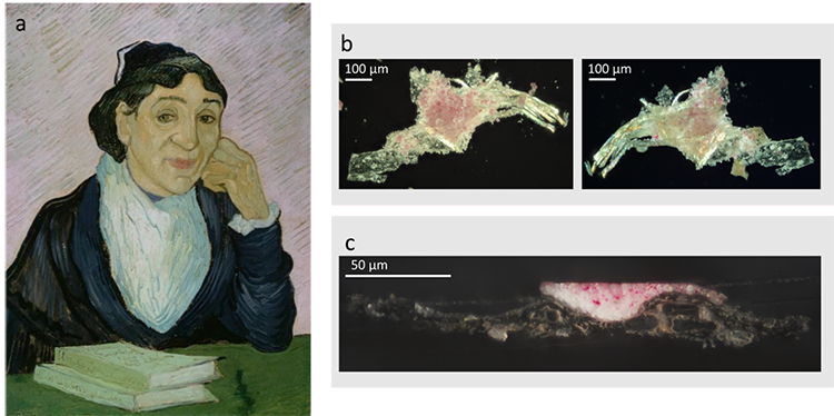
a) L’Arlésienne (portrait of Madame Ginoux) from Van Gogh, 1890. Kröller-Müller Museum
b) fragment extracted from the painting
c) thin section of the fragment.
OPTIR spectroscopy is a recently developed technique which exploits the subtle expansion of the materials when they are exposed to infrared radiation providing molecular information on the chemical compounds present and how they are distributed across the sample with a nanoscale resolution. In the context of historical paintings, OPTIR allows the characterization of nanometric particles and very thin layers. Nonetheless, compared to conventional infrared microspectroscopy coupled to synchrotron radiation (SR-µFTIR), the spectra obtained by OPTIR are noisier and, due to their high resolution, are also much slower. Thirdly, there is a possibility of photodamage in OPTIR due to the high power density of pump and probe lasers.
For this reason, the combination of OPTIR with SR-µFTIR proved to be an optimal method for the characterization of the samples. SR-µFTIR provided high quality spectra (high signal-to-noise ratio, accurate peak position, and flat baseline), non-destructive and fast measurements that allowed the characterization of the full sample, while OPTIR demonstrated to be a powerful technique for the analysis of submicrometric sample features. Indeed, the application of this multi-technique approach in a small fragment of the painting L’Arlésienne (portrait of Madame Ginoux) from Van Gogh provided detailed information on the artwork’s composition.
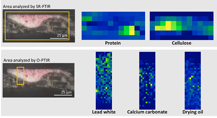
Top: results obtained by SR-µFTIR (pixel size 6 µm x 6 µm).
Bottom: results obtained by O-PTIR (pixel size 0.45 µm x 0.45 µm). The orange rectangles are 88 µm x 40 µm for the SR-µFTIR map and 4.5 µm x 17.5 µm in the OPTIR map.
SR-µFTIR analysis showed the presence of thick layers of protein and cellulose at the bottom of the sample, corresponding to the support of the painting, in this case a canvas made of cellulose fibers coated with a sizing. Results also show the presence of drying oil, lead white and calcium carbonate in the painting layer. In a complementary way, the analysis of the same sample by O-PTIR revealed the distribution of the pigments within the painting layers, showing the heterogeneous dissemination of lead white and calcium carbonate. The presence of geranium lake particles, probably associated to the red color observed in the background, was only detected thanks to the higher resolution of the OPTIR technique.
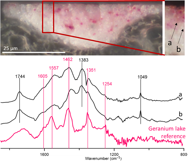
Top: region selected for the analysis (left) and its magnification with the analyzed points in evidence, a-b (right).
Bottom: O-PTIR spectra collected at each point and reference spectrum of geranium lake obtained by the same technique. The peaks marked in pink correspond to geranium lakes.
Geranium lakes have been previously observed in other paintings by Van Gogh. The use of this family of pigments is normally deduced from the detection of bromine in pink colored areas; however, the identification of the pigment molecules is difficult since they are normally mixed with drying oil containing similar functional groups, namely carboxyls, carbonyls and alkene bonds. Since geranium lakes tend to fade when they are exposed to light, its identification is very important to decide the best conservation conditions of this artwork.
These results highlight the possibilities of this novel analytical approach, which has the potential to become a fundamental tool in the analysis of cultural heritage.