SMIS
SMIS is an infrared spectromicroscopy beamline that delivers high-brilliance radiation in the mid and far-IR spectral range. The synchrotron beam is split to operate two endstations in parallel. The beamline is dedicated to multiscale spectromicroscopy, allowing up to ~20 nm spatial resolution and up to several cm sample size combining several instruments. We offer a variety of environmental conditions including pressure, temperature, atmosphere and polarization control.
Team

Beamline news
All SMIS news
European School HERCULES 2026 at SOLEIL
SOLEIL's contribution to the biology community - Scientific Meeting, Montpellier, April 28 (...)
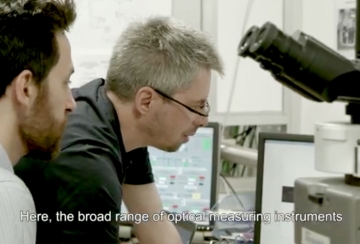
Videos
All SMIS Videos
The dust messengers
Hair in the spotlight
Beamline Specification
- Spectral range
-
8000 cm-1 – 200 cm-1 | 1.25 μm – 50 μm | 1 – 0.025 eV
- Spectral Resolution
-
0.1 cm-1
- Source
-
Edge and Bending Magnet Radiation - 78 mrad Horizontal × 20 mrad Vertical extraction
- Flux @ first optical element
-
1.86×10¹⁴ Photons/s/0.1%bw @ 10 µm for 500 mA stored current
- List of techniques
-
Single point microscopy
Time resolved, high spatial resolution microscopy (rapid scan and Step/Scan)
Single point raster mapping
ATR microscopy in inverted geometry
Very high sensitivity ATR spectroscopy (~70 ATR bounces)
2D IR FPA imaging
nanoIR techniques: sSNOM and phototermal AFM-IR (under commissioning)
mIRage photothermal IR microscope with ~500 nm resolution (operating with QCL source)
Raman microscopy
Müller polarimetry (SR adaptation in progress, available with thermal source)ATR and grazing incidence objectives
Diamond anvil cells: low (0.5-5 GPa) and moderately high (<40GPa) pressure
Cooling and heating sample stages (Linkam)
LHe flow cryostat (Oxford Microstat)
- Beam size at sample
-
Diffraction limited, up to ~5×5 μm2
- Flux on sample
-
~4.8×10¹³ Photons/s/0.1%bw at the entrance of the interferometers
- Detectors
-
small area MCT/A (Mercury Cadmium Telluride), MCT/B detector with extended spectral range, LHe cooled CuGe, InSb, Si photodiode
- Polarization
-
Linearly (bending magnet emission) and radially polarized (edge radiation)
Scientific opportunities
| Soft matter | Microscopic Analysis of inclusions in polymers; polymer interfaces; chemical imaging of components and crystallinity;deformation under stretching;Chemical composition changes with temperature |
|---|---|
| Geology | Identification of inclusions in minerals; Chemical imaging of inclusions constituents; Interface studies, grain boundaries between minerals; archaeological minerals; Study of water recirculation in deep earth mantle; Deformation and chemical composition changes of minerals under high pressure |
| Biology, Biomedical | Individual cells study, Imaging biochemical compositon inside individual cells: Apoptosis et Necrosis; Tumoral cells; Study of human tissues: hair, skin, bone, brain... ; bacterial identification. |
| Archeology | Analysis of ancient mummies, ancient cosmetics; analysis of traces of paint composition. |
| Thin Films | Analysis and chemical imaging of thin films, protection layers |
| Applied Research | Analysis and expertise of various applied problems in a short time scale. |
Desription of experimental capabilities at the SMIS beamline both with synchrotron beam and off line instruments.
Endstations
- Bruker Hyperion II ILIM / Invenio R
-
Location SMIS 1 Spectral range MCT detector (750 - 6000 cm-1), FPA imaging detector (900-2000 cm-1) Spectral resolution 0.125 - 32 cm-1 Operation modes Confocal dual aperture, single aperture, reflection and transmission geometry Accessories High quality purge box, microfluidic devices, large scale mirco-ATR accessory Objectives 15x (0.4 NA, WD: 24 mm), 36x (0.5 NA, WD: 10.4 mm) Sources Synchrotron source (300-8000 cm-1), QCL source (1000-1800 cm-1), Globar source
- Attocube/NeaSpec NeaScope s-SNOM/PTIR
-
Location SMIS 1 Spectral range MCT detector, 650 - 6000 cm-1 Spectral resolution 4 - 32 cm-1 Spatial resolution <20 nm Operation modes scattering SNOM spectromicroscopy, imaging with a QCL source and at 532 nm; photothermal AFM-IR spectroscopy and imaging with QCL source Sources Synchrotron source (300-8000 cm-1), QCL source (900-1850 cm-1), 532 nm laser
- Continuum microscope / Thermo Nicolet 8700 bench
-
Location SMIS 2 Spectral range MCT/A detector, 650 - 6000 cm-1
MCT/B detector, 400 - 6000 cm-1
Spectral resolution 0.125 - 32 cm-1 Operation modes Confocal dual aperture, single aperture, reflection and transmission geometry Accessories Linkam heating stage, microfluidic devices, Thermo ATR accessory Objectives 15x (0.58 NA), 32x (0.65 NA), 15x ATR ZnSe Sources Synchrotron source (100-8000 cm-1), Globar source, NIR Source The Thermo Nicolet 8700 bench is capable of time resolved studies in Rapid Scan and Step/Scan modes.
- Nicplan microscope / Thermo Fischer iS50 bench
-
Location SMIS 2 Spectral range MCT/A detector, 650 - 6000 cm-1
Si Bolometer, 100 - 900 cm-1
Spectral resolution 0.125 - 32 cm-1 Operation modes Confocal dual aperture, single aperture, reflection and transmission geometry Accessories Linkam heating stage, microfluidic devices, Diamond Anvil Cells Objectives 15x (0.58 NA), 32x (0.65 NA), Bruker Grazing Incidence Objective Sources Synchrotron source (100-8000 cm-1), Globar source, NIR source
- Horizontal microscope / Thermo Fischer iS50 bench
-
Location SMIS 2 Spectral range MCT/A detector, 650 - 6000 cm-1
Si Bolometer, 100 - 900 cm-1
Spectral resolution 0.125 - 32 cm-1 Operation modes Reflection and transmission geometry Accessories Diamond Anvil Cells (DAC), DAC LHe flow cryostat (base T~15K), DAC heating accessory (T up to 700 Cº), custom user experiments Objectives 15x (0.5 NA) Sources Synchrotron source (100-8000 cm-1), Globar source, NIR source
- mIRage photothermal IR and simultaneous Raman microscope
-
Location SMIS extension Spectral resolution 2 cm-1 Operation modes Reflection (Si and APD detectors for high and low power operation), Transmission (Si detector) Accessories Raman spectrometer (in progress) Sources QCL source (1000-1800; 2600-3050 cm-1), 532 nm laser
- Continuum XL microscope / Thermo Nicolet 6700 bench
-
Location SMIS extension Spectral range MCT/A detector, 650 - 6000 cm-1 Spectral resolution 0.125 - 32 cm-1 Operation modes Confocal dual aperture, single aperture, reflection and transmission geometry Accessories Linkam heating stage, microfluidic devices, Thermo ATR accessory Objectives 15x (0.58 NA), 32x (0.65 NA), 15x ATR ZnSe Sources Globar source, NIR source
- Agilent Cary 630/690 FTIR imaging system
-
Location SMIS extension Spectral range MCT detector, 750 - 6000 cm-1
FPA detector, 128x128 pixel, 900-3600 cm-1
Spectral resolution 0.125 - 32 cm-1 Operation modes Confocal dual aperture, single aperture, reflection and transmission geometry Accessories Linkam heating stage, microfluidic devices, Agilent µATR accessory Objectives 15x (0.58 NA), 25x (0.65 NA) Sources Globar source
- Müller Polarimeter / Thermo Nicolet 8700 bench
-
Location SMIS extension Spectral range MCT detector, 750 - 6000 cm-1 Spectral resolution 0.125 - 32 cm-1 Operation modes Reflection and transmission geometry Focus spot ~2 mm Sources Globar source
SMIS, in collaboration with multiple research groups is participating in the development of Quasar, an open source project, a collection of data analysis toolboxes extending the Orange suite. We empower researchers from a variety of fields to gain better insight into their data through interactive data visualization, powerful machine learning methods and combining different datasets in easy to understand visual workflows.
Potential users can download the software from the Quasar website and interested developers can contribute to the repositories of the Quasars GitHub organization.

Ruby fluorescence
References
Pressure dependence of ruby shift:
Shen G., et al. High Pres. Res. 40, 299 (2020)
Dewaele A., et al. Phys. Rev. B 78, 104102 (2008)
Mao H.K., et al. J. Geophys. Res. 91, 4673 (1986)
Temperature correction:
Datchi F., et al. High Press. Res. 27, No 4, 447-463 (2007)
Rekhi S., et al. High Temp. - High Pres. 31, 299 (1999) - for T > 298