Cyanobacteria intracellular biomineralization: Back in time with bacteria
Cyanobacteria are abundant bacteria in many continental and marine environments. 2.3 billion years ago they "invented" oxygenic photosynthesis process, this essential reaction that uses light energy to convert water and carbon dioxide into organic carbon and dioxygen. Thus, they have deeply impacted the chemical processes at the surface of the Earth since the advent of organic life. They have also played a major role in the formation of limestones by biomineralization. However up to day, there is still a poor understanding of how these bacteria catalyze this biomineralization and how this process evolved over geological timescales.
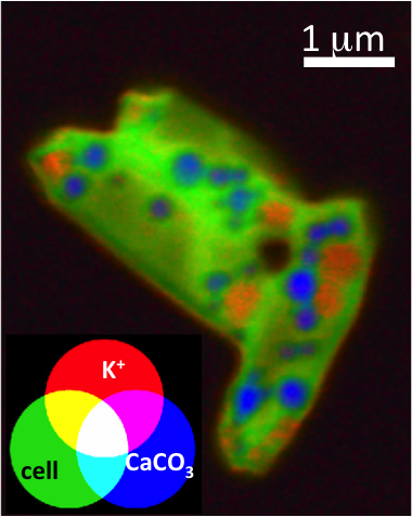
The recent discovery of some cyanobacterial species that are capable of forming amorphous calcium carbonates inside their own cells has radically changed our understanding of this process. How these cells regulate calcium is likely a discriminative feature with respect to other species of cyanobacteria incapable of such intracellular biomineralization.
On HERMES beamline, the STXM (Scanning transmission X-ray microscopy) spectromicroscopy setup, now available to users, offers key tools to identify and quantify the different intracellular reservoirs of calcium in cells and their variations. This allows a better understanding of the nucleation and growth mechanisms of intracellular CaCO3 in cyanobacteria and how the cells control them. Moreover, this approach will more generally open up new perspectives and opportunities for studying the regulation of calcium by cells.