A better decoding of nanoparticle surfaces at the atomic scale
A research team has developed a method that allows free nanoparticles to be studied while avoiding any substrate influence. In this way it becomes possible to specifically characterize the surfaces of nanoparticles. Prospects are numerous, in particular concerning functionalized nanoparticles, the applications of which are very promising in the biomedical and energy fields. The study is published in the Journal of Physical Chemistry Letters.
Conventional X-Ray photoemission spectroscopy (XPS) is the most common technique used to study the surfaces of materials, yielding information on the chemical composition of a thin layer, just a few dozen atoms thick. Until now, in order to study the surface of nanoparticles, it was necessary to deposit them on a substrate. However, interactions between the nanoparticles and the substrate, coupled with charge effects on the sample, make such XPS data difficult to interpret.
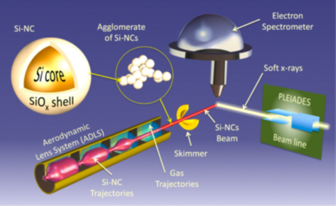
A team from the CEA IRAMIS laboratory, the SOLEIL synchrotron, the Institut Lavoisier de Versailles (UVSQ/CNRS) and the Institute of Physics · Rennes has recently developed a new experimental method of tackling this problem. With their apparatus, the interaction between the nanoparticles, well defined both spatially and temporally by means of an aerodynamic lens, and a beam of X-rays, can be studied. The experiments were performed, under high vacuum conditions at the PLEIADES beamline (at SOLEIL synchrotron).
In a demonstration experiment, particles used to demonstrate the feasibility of the method were silicon nanocrystals with about 14 nm diameter (1nm= 1 billionth of a meter). They were synthesized using laser pyrolysis at CEA IRAMIS, and oxidized in ambient air prior to measurements at the synchrotron. The analysis of data collected at PLEIADES, showed a continuous transition of the interface pure silicon / oxidized silicon for a thickness of less than 1 nm.
Previous scientific studies on similar objects had been performed previously with samples deposited on a substrate. However, the interaction with the substrate did not allow the origin of the observed phenomena to be well elucidated. The new method allows effects related only to the nanostructure of matter to be distinguished, thus avoiding the parasitic influence of the substrate.
In addition, this new technique is extremely sensitive to any modification occurring on the surface of nanoparticles in interaction with several chemical environments. It will be applicable to the characterization of a broad range of nano-objects, in particular, functionalized systems where active molecules of various complexity, are “anchored” to the nanoparticle surface. Because of the particular structure of matter at the nano-scale, the functionalized nanoparticles have astonishing physical and optical properties, with many applications for example in photoluminescence imaging or in treatment of tumors with targeted radio and phototherapy.