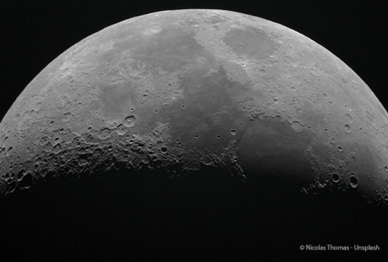
Urinary lithiasis (renal calculi) is a common disorder which is becoming more widespread in all industrialized countries. Several different pathologies are grouped together under the general term “renal calculi”, some of which, notably hereditary lithiasis, can evolve into terminal renal insufficiency without early diagnosis and appropriate treatment. It is necessary to know the cause of the disease in order to treat it efficiently, but unfortunately the diagnosis of this type of lithiasis is often identified lately, thus reducing the chances of saving the patient’s kidneys and, in the case of terminal renal insufficiency, presenting a proven risk of transplant failure.
The challenge was therefore to carry out an early diagnosis of the disease. The experimental procedure was already tested at the infrared microspectroscopy SMIS beamline at SOLEIL . Based upon several months of previous study the expected results have been obtained in just a few hours.
Dominique Bazin, a research scientist at the “Laboratoire de Physique du Solide” and Dr Michel Daudon from the Necker Hospital screened ,using this infrared light ,several biopsy samples from the kidney of a patient hospitalized for severe renal insufficiency of unknown origin. They looked for signs of the presence of crystalline deposits in order to identify which of three possible causes was the reason for the severe renal insufficiency of this particular patient at the Necker Hospital. The initial clinical diagnosis suggested adapting the analyses to a search for the presence of 2,8-dihydroxyadenine (DHAd), a compound produced by the body when it has difficulty-metabolizing adenine, a molecule that plays a vital role in human beings.
The very small size of the crystals studied (a few microns) together with the high precision required to identify DHAd within the tissue were severe constraints that the SMIS group is already used to.
Quickly, the presence of DHAd crystals has been identified on the samples, resulting in a quick personalization of the treatment for this patient as a consequence. An early diagnosis that made all the difference, as the medical treatment of the disease thus identified was available and could be applied at once.

| Chemical mapping of DHAd created with an aperture of 6x6 microns, allowing the nature of the inclusions in the kidney section to be identified |
The presence of DHAd crystals has been identified
Dominique Bazin, a research scientist at the “Laboratoire de Physique du Solide” and Dr Michel Daudon from the Necker Hospital screened ,using this infrared light ,several biopsy samples from the kidney of a patient hospitalized for severe renal insufficiency of unknown origin. They looked for signs of the presence of crystalline deposits in order to identify which of three possible causes was the reason for the severe renal insufficiency of this particular patient at the Necker Hospital. The initial clinical diagnosis suggested adapting the analyses to a search for the presence of 2,8-dihydroxyadenine (DHAd), a compound produced by the body when it has difficulty-metabolizing adenine, a molecule that plays a vital role in human beings.
The very small size of the crystals studied (a few microns) together with the high precision required to identify DHAd within the tissue were severe constraints that the SMIS group is already used to.
Quickly, the presence of DHAd crystals has been identified on the samples, resulting in a quick personalization of the treatment for this patient as a consequence. An early diagnosis that made all the difference, as the medical treatment of the disease thus identified was available and could be applied at once.