What mechanisms are responsible for the preservation of archaeological textiles over thousands of years?
An interdisciplinary team has revealed the mechanisms behind the exceptional preservation of 4,000-year-old textile remains from the Ancient East. The imaging of these archaeological samples in the different energy domains of the light spectrum made it possible to reconstruct a physico-chemical scenario explaining their morphological preservation at the nanoscale. More specifically, the X-ray microtomography (µ-CT) results obtained on the ANATOMIX beamline have made it possible to understand the key silicification (i.e., the replacement of organic constituents by silicate minerals) stage through the spatial speciation of the organic and mineral phases present in these heterogeneous specimens.
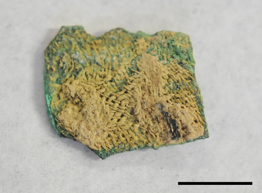
In archaeology, fragments of ancient textiles can be particularly valuable: in some cases, they provide us with the only accessible insight into ancient textile production for clothing or ritual purposes, but also for many technical functions such as the transport of materials, packaging or the production of objects. Unfortunately, organic textiles, composed mainly of cellulose, tend to degrade very quickly in the environment and few of these artifacts survive over the ages. However, as early as the 19th century, archaeologists excavating major sites in the Middle East were surprised by the existence of a number of identifiable textile imprints found petrified on the surface of objects, often metallic (FIG. 1).
This phenomenon, referred to as "mineralization", had already been observed even in conditions that were highly detrimental to the preservation of organic matter. By 1751, a definition of "petrification" had already been included in the Encyclopédie ou Dictionnaire raisonné des sciences, des arts et des métiers edited by Denis Diderot and Jean Le Rond d'Alembert. With the help of X-ray microtomography (µ-CT), this "natural process whereby a body from the plant or animal kingdom is converted into stone whilst preserving its original shape" has now been described at a sub-microscale thanks to experiments conducted at SOLEIL
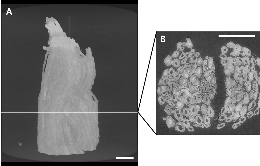
Cellulosic fibres collected from exceptionally preserved textiles and exhumed in the vicinity of copper objects were imaged by µ-CT on the ANATOMIX beamline. The 2D or 3D virtual sections and volumes (FIG. 2), with a spatial resolution of less than 1 µm, made it possible to determine the histology of the fibres as well as certain alterations such as micro-cracks.
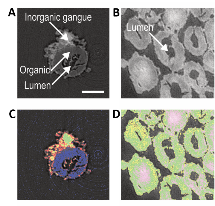
By implementing a semi-quantitative protocol for microtomography data acquisition and analysis, the grey levels of the images could be correlated to different organic and mineral phases (FIG. 3). A "positive mineralization" could be demonstrated for the most mineralized fibres via the presence of siliceous phases in all their cell walls (FIG. 3D).
(A and B) Virtual sections associated with a sparsely mineralized fibre from the Gonur-Depe site in Turkmenistan
(A) and highly mineralized fibres from the Tello site in Iraq (B).
(C and D) Images obtained by segmentation of virtual sections showing the distribution of the main phases present (blue, organic phase; red, phase composed of soluble calcium, silicon, and copper compounds; yellow, green and purple, copper silicates). Scale bar: 20 µm.
Combined with atomic force microscopy–infrared (AFM-IR) and second harmonic generation (SHG) microscopy, the µ-CT results provide a better understanding of the processes that led to the exceptional preservation of cellulosic materials at the sub-microscale. For a short period of time after burial of the textiles, the fibres are protected from rapid microorganism degradation by the biocidal action of metal cations released during the corrosion of nearby buried metal objects. Year after year, the cellulose chains are chemically degraded and the cellulose macrofibrils lose their crystallinity. This disorganisation in turn encourages the dispersion of solutes from the soil, first on the surface of the fibres, then more slowly through their cell walls. Eventually, this results in their silicification by condensation of silicate compounds in the manner of sol-gel syntheses used in chemistry, which gives them a mineral or "petrified" appearance.
This study is ongoing at the Supramolecular and Macromolecular Photophysics and Photochemistry Laboratory (PPSM, ENS Paris-Saclay, CNRS), on the basis of the study of a large corpus of Celtic textiles from the Iron Age at a sub-microscale, as part of Clémence Iacconi's thesis research, which is conducted on SOLEIL's PSICHÉ and PUMA beamlines in collaboration with a large research consortium involving French and Dutch teams.