Synchrotron SOLEIL actively participates in SARS-CoV-2 research
In the combat against the SARS-CoV-2 virus, the French National Research Agency (ANR) launched a call for projects at the beginning of March 2020, for the funding of targeted research topics on the study of the virus. In a concerted effort to advance our understanding of this coronavirus, scientists from SOLEIL's "HelioBio" Biology/Health section (PROXIMA-1 and DISCO beamlines) have joined forces with INRAe and Sanofi in a joint research project referred to as AcceS-Ge CoViD-19, which aims at determining the crystallographic structures of the virus proteins. At the end of March, out of 270 submitted research proposals, the ANR selected 44 projects including the project submitted by Synchrotron SOLEIL, for immediate funding.
Project introduction
The first step is to clone all the virus proteins responsible for CoViD-19. Cloning involves isolating the piece of DNA that contains the necessary information to produce a given protein, and inserting it into a "mini-factory" to produce this specific protein in large quantities. The mini-factory in question is usually a bacterium or eukaryotic cell (yeast, plant or mammalian cell...). The aim of the project is to carry out this operation for each of the SARS-CoV2 proteins. The researchers will then work on either the whole proteins or just their "interesting" parts - for example, the virus envelope protein that interacts with a target cell in the infected person. Scientists will then apply accelerated structural genomics to these different proteins or protein fragments in an internal platform within Synchrotron SOLEIL.
More specifically, the cloning step consists in manipulating isolated fragments of the virus' DNA in order to produce inactive viral proteins, which are harmless on their own as they are separated from other viral proteins. In classical structural biology techniques, it is common to clone the DNA of the protein being studied in order to have it produced by bacteria into which the DNA fragment carrying the information for this protein will have been inserted. These proteins are then purified through classical methods and subjected to crystallisation screenings. In other words, dozens, even hundreds of different conditions are tested in an automated process (using various types of solvent, various concentrations of protein, etc.). Ultimately, the scientists aim to obtain the protein in the form of crystals, which are then analysed by X-ray diffraction on biocrystallography beamlines such as PROXIMA-1 and PROXIMA-2A at Synchrotron SOLEIL, and will provide access in an extremely-accurate manner to the 3D structure of this protein downs to atomic scales. Despite advances in technology and process automation, however, these purification and screening steps are often very time-consuming.
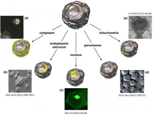
The objective of the platform is as follows: the protein of interest will not be produced then purified and crystallized through lengthy steps with a sometimes random outcome. It is during the actual protein manufacturing stage, within the overexpressing cells, which in this case is carried out by human cells rather than bacteria, that the screening takes place: different methods of inserting the DNA fragment of the protein into the cells, different cell culture conditions, etc., are tested. In certain "ideal" cases, the protein will be produced in exceptionally large quantities and will naturally form crystals, inside the manufacturing cells. Using this method, the purification and screening steps are avoided, greatly accelerating the process while remaining in a confined system. Researchers must then identify which of all the cells contain protein crystals – at this stage, the DISCO beamline comes in.
Following their production-crystallisation, by analysing the cells containing the protein crystals directly under an X-ray beam, the 3D structure of these proteins shall be identified on crystallography beamlines, such as PROXIMA-1. Once these structures have been unravelled, they will ultimately serve as targets for drug design studies. These advances should lead to a better understanding of the virus and its pathogenicity, and initiate the development of antiviral drugs.
Update on the development of the research project
As soon as the projects selected by the ANR were announced, the SOLEIL - INRAe - Sanofi team set to work, starting with the scheduling of the various deadlines. The challenge of obtaining the full pathogen genome in the context of lock-down proved to be successful in early May, which made it possible to initiate cloning of the SARS-CoV-2 proteins. To date, more than 30 constructs have been cloned. The next step is to transfect them into cells before in vivo crystallisation screening is conducted. To follow the step by step evolution of this project, visit the dedicated page