Rare earth elements are trapped inside the vessels of the conductive tissues of an accumulating fern
Rare earth elements (REEs) are strategic metals strongly involved in low-carbon energy conversion. However, these emerging contaminants are increasingly disseminated into ecosystems, raising concern regarding their toxicity. It is therefore important to better understand REE transfer to the trophic chain. In this study, we deciphered REE accumulation sites in the REE-accumulating model fern Dryopteris erythrosora by X-ray µfluorescence. REEs were translocated from the roots to the fronds by the xylem sap and were stored within the xylem vessels.
According to their atomic mass, the 17 rare earth elements (REEs) can be split into light REEs (LREEs from lanthanum to europium, plus scandium) and heavy REEs (HREEs, from gadolinium to lutetium, plus yttrium). Their physicochemical properties make REEs essential for numerous domains, such as green energy, industry, high technologies, and medicine. However, REE mining, intense utilization and low recyclability, are responsible for an increasing contamination of soils and waters. Phytoremediation is a nature-based solution consisting in the use of plants to remediate contaminated sites. Unraveling the transfer and storage mechanisms of REEs in plants is of utmost importance to understand metal transfer to the biota and to select the best candidates for phytoremediation.
Few plant species accumulate REEs. Among them, several fern species have been identified. The distribution of REEs in the chloroplasts or cell walls of tropical accumulating ferns was first reported either by tissue fractionation techniques or using radioactive tracers. However, experimental biases associated with tissue fractionation, loss of root integrity during REE radioactive tracer use, or indirect REE detection urged more accurate, nondestructive, and direct detection analysis provided by synchrotron μXRF. Moreover, the localization of HREEs in undisturbed tissues has not been performed previously.
In this study, resulting from a collaboration between the LUCIA beamline team, the P06 beamline team (DESY Synchrotron, Hamburg), the Laboratory Chrono-environnement (Univ. de Franche-Comté, Montbéliard) and LIEC (Univ. de Lorraine, Nancy & Metz), we analyzed the REE accumulation sites in D. erythrosora. This model fern species is cold resistant and was therefore chosen here to get knowledge on REE transfer in this promising species in the perspective of its further use in the phytoremediation of REE-contaminated sites in temperate/cold regions.
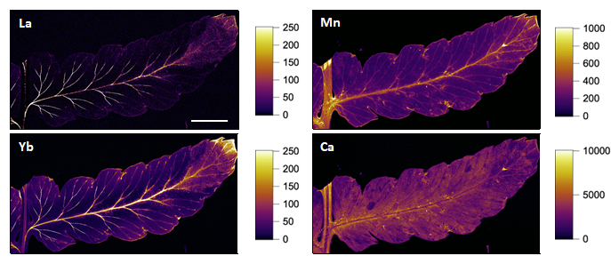
A first set of experiments at P06 allowed to resolve the REE distribution at the whole organ level. Both LREEs and HREEs were found in the conductive veins of the pinnae (sub-part of the frond) (Figure 1), a distribution different to that of nutrients such as K, and Ca.
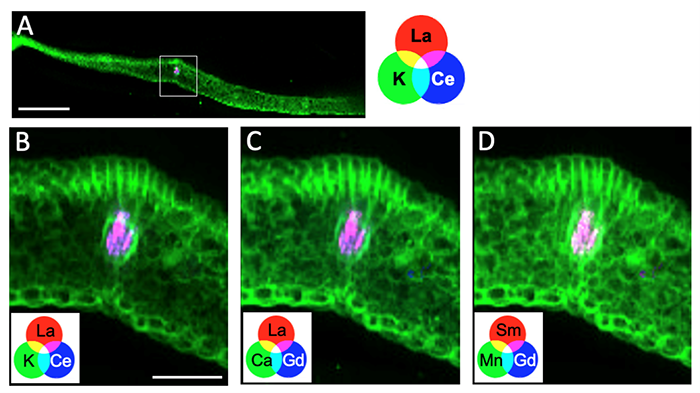
The accumulation of REEs in the vasculature prompted us to investigate in more detail the REE distribution by elemental μXRF mapping on thin cryosections of pinnae and rachis at LUCIA. REEs were distributed in the central vein rather than in the surrounding cells (Figure 2), which could not be inferred from Figure 1.
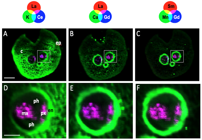
In the rachis of the frond, REEs were also found inside the conductive tissues as shown by the detection of REEs inside the vascular bundles (Figure 3). Conversely, K, Mn and Ca, were present mainly in the endodermis, cortex, and epidermis.
In conclusion, this study revealed that the REE distribution is associated mostly with conductive vessels in D. erythrosora. REEs could be trapped in the vessels due to their immobilization by ligands in the sap or to the lack of systems allowing their unloading out of the vessels. The comparison of REE cellular localization with elements that have contrasted redistribution/mobility potentials, such as Ca, Mn, and K, provided insights about the REE mobility in plants. Future investigations will focus on the comparison of REE storage sites among diverse plant species.