LUCIA
LUCIA is characterized by the production of a microfocused beam in the 0.8-8 keV energy range. The spatial stability of the beam spot over a wide spectral range enables elemental distribution studies by micro-fluorescence X-ray spectroscopy (µ-XRF) as well as elemental speciation by X-ray absorption spectroscopy (XANES and EXAFS) in heterogeneous samples.
The beamline "LUCIA" (Line for Ultimate Characterisation by Imaging and Absorption) is a "tender" (0.8-8 keV) X-ray microprobe with capabilities for chemical speciation by x-ray absorption spectroscopy (µ-XAS) and for elemental mapping by x-ray micro-fluorescence (µ-XRF). It allows the possibility to measure heterogeneous samples at a micronic size and to combine these two element-specific and non-destructive techniques.
A monochromatic beam of µm size is incident on a sample which is carried on a scanning x-z stage. µ XRF shows the location of the elements, their relative abundances, and with which other elements they are associated. One can take advantage of the monochromatic beam which allows to separate out different elements by their absorption edges. After a cartography of the sample by fluorescence, interesting spots can be analyzed by XAS to determine the speciation (local chemistry, quantitative determination of the local geometric structure around the absorbing atom) of the elements and how this depends on the different components.
µ-XRF and µ-XAS can be combined with other microtechniques like Raman spectroscopy to give complementary informations on the sample. The energy range offered by the beamline corresponds to the best performances of SLS and SOLEIL in terms of brilliance. It allows XAS experiments at the K edge of elements from Na to Fe, L edges from Ni to Gd, and M edges of rare earths and actinides.
Contacts
| Member | Scientifique expertise | Contact |
| Delphine Vantelon, beamline manager | Earth and soil sciences | +33 (0)1 69 35 96 94 |
| Nicolas Trcera, beamline scientist | Materials and surface sciences | +33 (0)1 69 35 81 23 |
| Solenn Reguer, beamline scientist | Cultural heritage | +33 (0)1 69 35 97 27 |
Camille Rivard, beamline scientist, INRAE associate engineer | Biology, earth and environmental sciences, | +33 (0)1 69 35 94 71 |
| Pierre Lagarde, emeritus beamline scientist | Materials and surface sciences | +33 (0)1 69 35 96 88 |
Beamline news
All LUCIA news
SOLEIL Highlights 2025
European School HERCULES 2026 at SOLEIL
Team

Videos
All Lucia Videos
Legacy phosphorus
The lights of SOLEIL
Technical data
- Energy range
-
0.6 – 8 keV
- Source
-
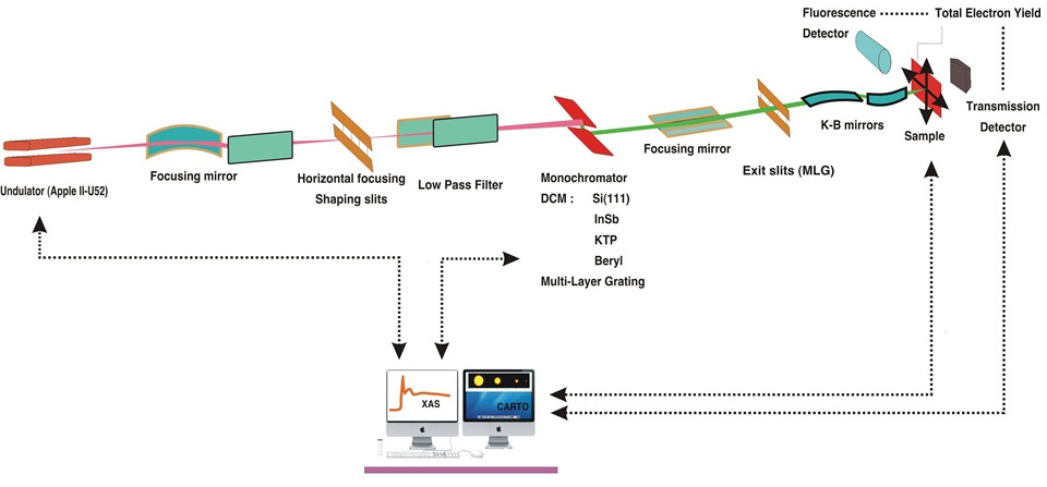
Undulator HU52 "Apple II" type, 32 periods, gap 15 – 150mm, variable linear polarization, left and right circular polarizations, operating on harmonics 3 to 21
- Optics
-
Spherical mirror for horizontal de-magnification; two planar mirrors act as a low pass filter to remove higher harmonics. Their angle of incidence can be varied from 0.4o to 1.3o. All mirrors are silicon coated with nickel.
- Monochromator
-
Double crystal monochromator built by Kohzu. Its design is based on a double cam which provides a fixed exit geometry and covers a large angular range (5o- 75o). Up to five different crystals, namely KTP(011), MGM (Multilayer Grating Monochromator), InSb(111), Si(311) and Si(111) fit into the water cooled holders.
- Energy resolution
-
A table describing the energy ranges and resolution for each monochromating system can be accessed
- Focusing optics
-
The final focusing is done with a "Kirkpatrick-Baez" (KB) reflecting mirrors system. It is based on an ESRF-design, and was modified to be high vacuum compatible.
- Spot size on the sample
-
3 x 2 mm² for the macro-beam
2.5*2.5 µm2 for the micro-beam
- Flux on the sample
-
8.0*1010 ph/s/400mA @ 4000eV in 2.5*2.5 µm2
1.6*1011 ph/s/400mA @ 4000eV in 1.5*1.5 mm2
- Sample environment
-
Measurements are always performed in a primary vacuum chamber (primary vacuum or secondary vacuum for cryogenic analyses or if the samples require it)
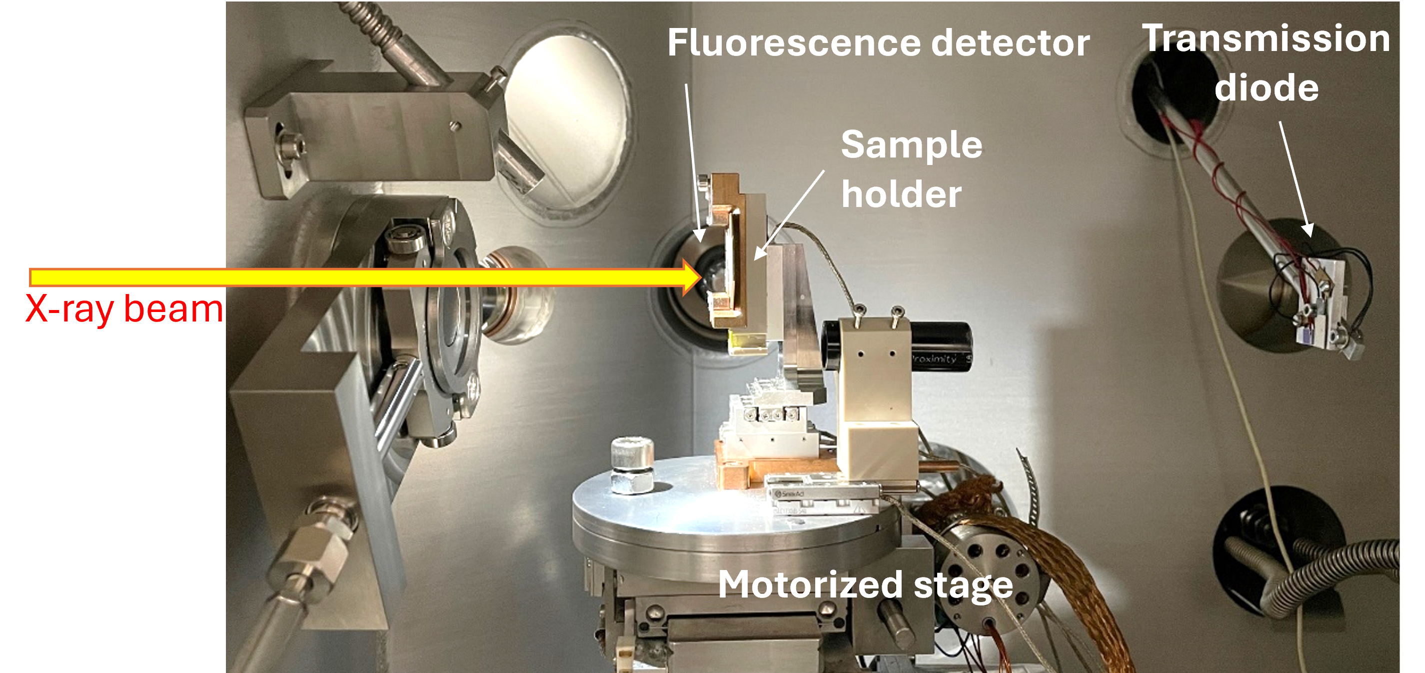
Chambre expérience de la ligne LUCIA. 2025 It is rather spacious and can accept a variety of setups. A motorized (x,y,z)-stage allowing the precise (µm) and reproducible (coders present) positioning of the sample for mapping purposes. The following environments can be added:
- A liquid / electrochemical cell
- An oven
- A He/N2 cryostat
- A peltier
Separate UHV chambers for surface science and chamber for electrochemistry are also available, they are mounted downstream of the main chamber.
- Detection
-
µXRF: measurement of the fluorescence spectra: SDD (silicon drift diode) 80 mm² Bruker
µXAS: measurement of the fluorescence yield (FY), of the sample drain current (TEY) and measurement of the transmitted photons flux (diode)
Sample holders
- Generalities
-
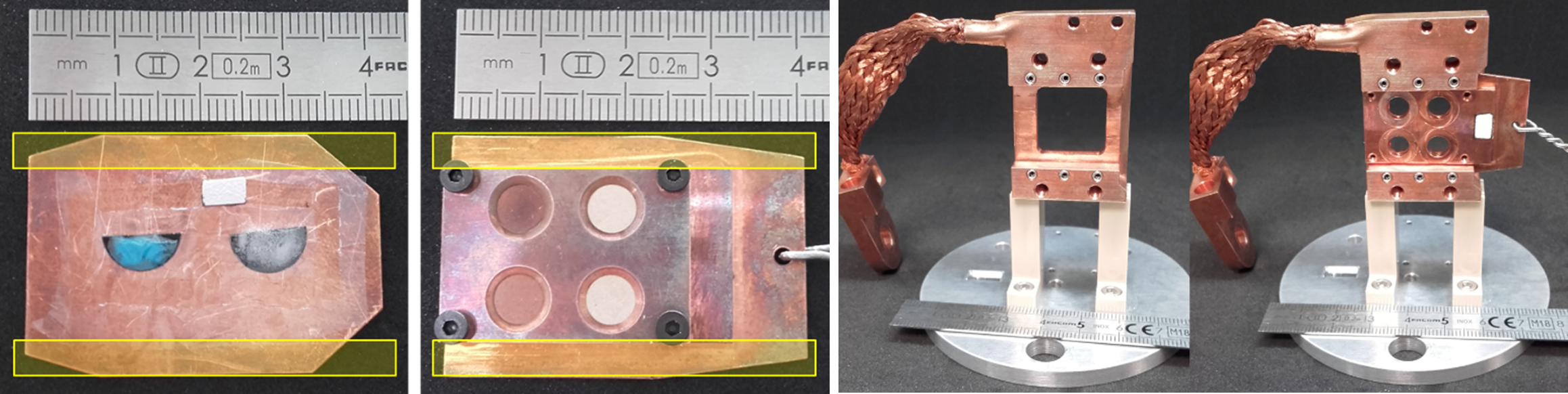
Most of the LUCIA sample holders are based on a copper plate put in two rails of the sample stage. The yellow area must be consequently left free. The copper plate is vertical in the sample stage.
Sample stage and classical sample holders on LUCIA
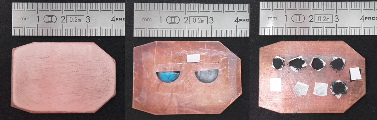
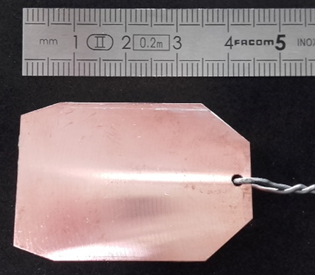
- Flat, simple Cu plate
-
Used to stick adhesive carbon tape with powder, to press indium foil with powder or to stick samples with adhesive double-sided tape. Ultralene foil must covered powered samples.
Detection allowed: fluorescence and TEY modes.
- Plate for pellets
-
Four 10 mm diameter pellets can be placed on the plate and a platelet is screwed using 4 screws (steel or molybdenum screws). All pellets mounted on a plate must have the same thickness.
Detection allowed: fluorescence, TEY and transmission modes.
Plate for pellets on LUCIA
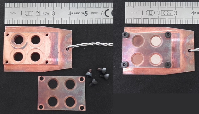
- Thick plate for “large” samples
-
Used for samples larger than 20 mm in diameter. There are still some sample size constraints in the three directions (maximum size is 5 cm both in vertical and horizontal). Check the size of your samples with your local contact.
Detection allowed: fluorescence and TEY modes.
Thick plate for “large” samples on LUCIA
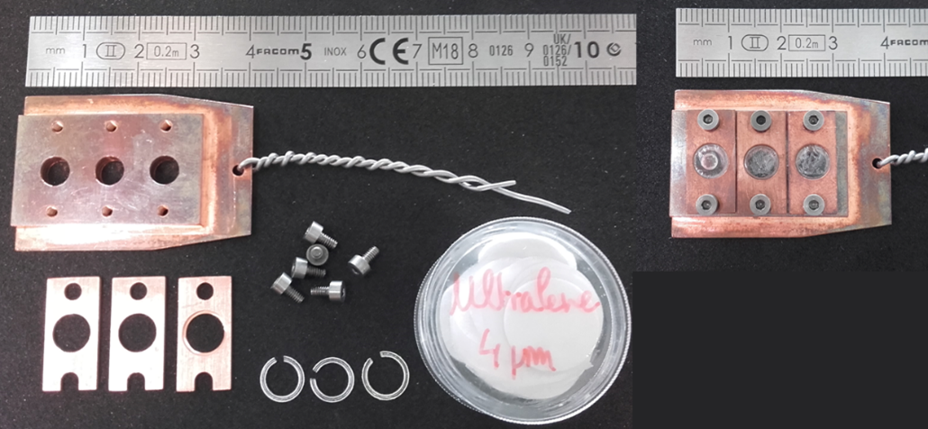
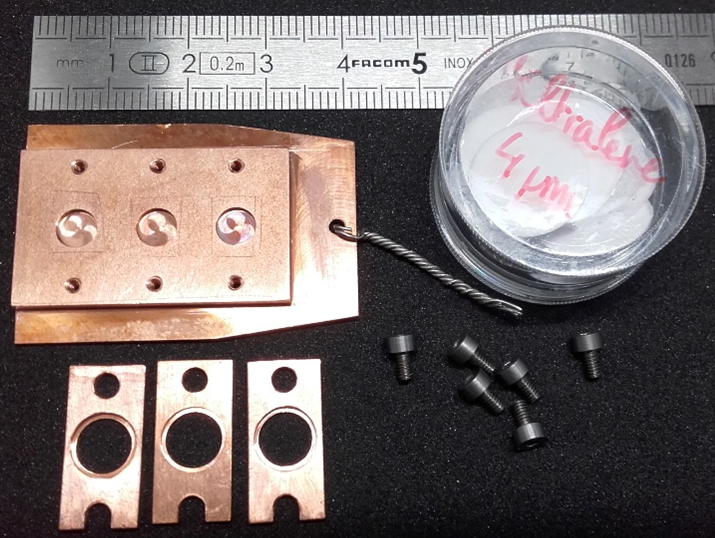
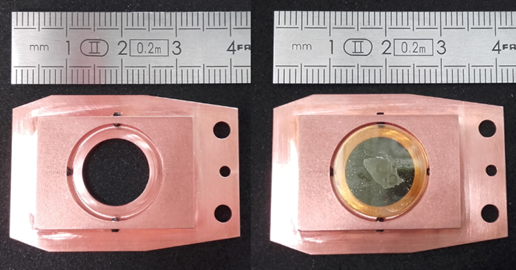
- Thick plate with holes for biological cryo thin sections
-
Three cryo thin sections can be sandwiched between two Ultralene foils and fixed with a ring system on Cu platelets. Cu platelets are screwed on Cu thick plate using two screws (steel or molybdenum screws). Diameter of the holes of the platelet is 6 mm and of the holes of the Cu thick plate is 5 mm.
Other type of thin samples can be also placed on Ultralene foil stick on PEEK platelets, screwed using two screws (steel or molybdenum screws). The square opening is 5 x 5 mm².
Detection allowed: fluorescence mode.
Thick plate with holes for biological cryo thin sections on LUCIA
- Thick plate with wells for frozen solution
-
Three solutions can be put in the wells of the sample holder. The volume of a well is 25 µL. The sample holder is plunged in liquid N2 to freeze the sample. Then, on the front of the sample holder, Cu platelets with Ultralene foil are screwed using two screws (steel or molybdenum screws).
Detection allowed: fluorescence mode.
Thick plate with wells for frozen solution
- Plate for 1 mm thick - 16 mm diameter slide
-
The slide is fixed on its sides by four screws. It was designed for a SMIS-LUCIA combination. The ZnSe disc inserted in the centre is compatible with certain analyses on SMIS.
Detection allowed: fluorescence mode.
Plate for 1 mm thick - 16 mm diameter slide
Scientific opportunities
| Surface Science | An UHV set-up is currently available on the beamline. It is equipped with a sample preparation chamber (furnace, evaporation cells, ion gun) and a data acquisition chamber allowing a sample characterization with LEED, Auger, XPS and x-ray spectroscopy measurements by total electron yield. In a next future, fluorescence yield will be also installed. Experiments on silicene deposited onto Ag(110) and Ag(111), silver clusters deposited on Al2O3(110), MgO on Ag(100) have already been conducted on the beamline (unpublished results). |
|---|---|
| Material Science | The characteristics of the LUCIA beamline are perfectly adapted for material sciences. It can take advantage of the µ-focused beam and the adaptable energy range for studying heterogeneous materials. Several equipment are available to probe matter under extreme conditions such as high pressure (diamond anvil cell) or high/low temperatures (ovens and cryostats). Moreover, particular setups can be simultaneously combined in the experimental endstation (Raman – XAS). A wide range of scientific areas can be investigated on LUCIA, from fundamental to applied sciences (technical materials, Earth science, Cultural heritage, Environment,...). |
| Chemistry | Several equipments can be adapted to the endstation of LUCIA in order to probe chemical reactions in situ. In particular, the study of liquids, as well as solid-liquid interfaces is possible. Liquid, electrochemical or catalysis cells are being developped and can be designed for specific needs. Scientific areas explored at LUCIA with these sample environments range from supported catalysis and thin film electrochemistry to elemental speciation in oil residues or nanoparticle synthesis routes. |
| Life Science | Elemental characterisation in animal or plant organs and tissues, cells: these studies aim to determine the distribution and track the fate of nutrients in plants (e.g. phosphorus), pollutants (metals, rare earths, nanoparticles) or metal ion transporters (metalloproteins) using X-ray fluorescence imaging. Coupling with µ-XANES analysis makes it possible to determine the speciation of elements directly within cells or tissues. To best preserve the structure and chemical composition of the sample, rapid freezing by immersion in isopentane cooled with liquid N2 is generally used. Cells can be analysed directly on their freezing medium (ultralene film, Si3N4 window), while organs and tissues are cut using a cryomicrotome. X-ray fluorescence mapping analyses are generally performed cryogenically using the liquid N2 cryostat. |
Beamline scheme
Proposal submission
Proposal submissions are done through the SUNSET, le portail des utilisateurs de SOLEIL.
The submission dates are Februay 15th and Septembre 15th.
It is strongly recommented to contact one of the beamline scientist (see Presentation/Contacts) before submitting a proposal.
Links
The x-ray data booklet for X-rays and other photons users edited by the Center for X-ray Optics and the Advanced Light Source.
Publications
Publication technique générale sur LUCIA:
- LUCIA, a microfocus soft XAS beamline (511.97 KB)
Publication sur l'outil de visualisation du micro-faisceau de LUCIA:
Publications sur le couplage µRaman - µXRF sur LUCIA:
- Combining two structural techniques on the micrometer scale: µXAS and µRaman spectroscopy (868.04 KB)
- Applications in materials science of combining Raman and X-rays at the macro and micrometric scale (1.38 MB)
Publications d'utilisateurs représentatives des possibilités offertes par LUCIA:
- Local structures around Na, K, Ca, Mn, Fe and Cu in medieval glasses: effect of weathering (92.45 KB)
- High pressure x-ray absorption at low energy (31.85 KB)
- Investigation on corrosion of ion archaelogical artefacts using microfocused synchrotron X ray absorption spectroscopy and imaging (146.67 KB)
- Microscale distribution and speciation of Pb and Sb in shooting range soils (107.16 KB)