Kidney biopsy studies: a combination of several techniques that improves their accuracy
A new step towards more accurate diagnosis for severe or acute renal failure just has been achieved. In a recent study published in Comptes Rendus Chemistry, scientists combined field effect scanning electron microscopy, deep UV fluorescence, Raman spectroscopy and both classical and synchrotron radiation Fourier Transform Infrared spectroscopy to take a special look to crystal-containing kidney biopsies.
This work has been conducted by scientists from the University Pierre and Marie Curie, the Laboratoire de Physique des Solides (Orsay, France), Inserm, the Imperial College of London, and the SOLEIL synchrotron facility. This set of method appears to successfully provide information regarding the nature and the spatial distribution at subcellular scale of different chemical phases present in kidney biopsies, as well as on the crystal morphology. The SMIS beamline synchrotron radiation allowed performing µFTIR non-destructive experiments when crystals were too small for classical FTIR microscopy.
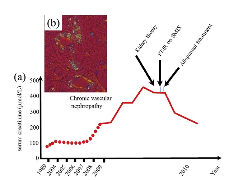
(a) Evolution of the kidney function as given by the serum creatinine. The medical diagnosis from crystals in the kidney biopsy cannot be performed at the hospital (in a short time). As we can see the FTIR measurements performed on the SMIS beamline of the Soleil synchrotron radiation center allow us to identify unambiguously the crystals and allow the clinician to give the right treatment and finally the serum creatinine decreased significantly avoiding dialysis and a kidney graft for the patient.
(b) Abnormal deposits observed in the tubular cells and interstitium.
All these techniques are already at the core of an early diagnosis tool kit and may allow clinicians to collect a wide range of information and to identify unambiguous crystal deposits involved in kidney dysfunction. Finally, the latest developments performed in UV visible micro-spectroscopy show that we are on the verge of collecting exciting results regarding the spatial distribution of organic compounds at a resolution reaching 150 nm. Pathological calcifications are not limited to kidney. Therefore, this set of diagnosis could benefit to a large range of microcrystalline pathologies.