Industrial anti-cancer research, assisted by diffraction data collected at SOLEIL, is reported in the journal Nature
The intrinsic apoptosis (programmed cell death) pathway responds to irreparable DNA damages in cells by marking them for destruction. Cancerous cells with damaged DNA distinguish from classical cells by mechanisms that avoid apoptosis, and consequently continue to proliferate even though they are not functioning normally.

The intrinsic apoptosis pathway is regulated by a series of pro-apoptotic and anti-apoptotic BCL2 proteins. Thepro-apoptotic BCL2 proteins BAK and BAX are able to aggregate and form channels in the outer membrane of the mitochondrion, allowing the release of cytochrome C into the cytoplasm, which leads to the activation of caspase enzymes and hence initiate the programmed cell death. In well-functioning cells, anti-apoptotic BCL2 proteins prevent channel-forming aggregation of BAK or BAX. When the cell is marked for death, for counter example as a result of irreparable DNA damage, a further set of pro-apoptotic BCL2 proteins are produced by the cell sequestering anti-apoptotic BCL2 proteins in the cytoplasm, and hence indirectly encourage the aggregation of BAX and BAK.
In many types of cancer (for example acute myeloid leukaemia or T-cell lymphomas) the balance of this regulation is shifted, with the cancer cells overproducing anti-apoptotic BCL2 proteins in order to avoid programmed cell death. These so-called « survival group » proteins are then targets for drug action – if the « apoptosis blockers » in cancerous cells can be sequestered (with minimal side effects on the « apoptosis blockers» in normally functioning cells), this provides a means of preferentially encouraging cancer cells to commit suicide.
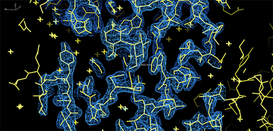
X-ray diffraction data, collected at SOLEIL’s PROXIMA-1 beamline, using industrial access beamtime purchased by the UK pharmaceutical company VERNALIS (and commissioned by research groups at SERVIER), have been published in the journal Nature. This study provided the structural information to help understand the function of the molecule S63845, which binds very strongly and specifically to the anti-apoptotic protein MCL1. Indeed several oncogenic pathways lead to MCL1 expression. The binding mode of S63845 (see Figure) mimics the action of anti- apoptotic proteins expressed in normally functioning cells. Initial tests indicate that S638345 has both low cytotoxicity and high efficacy against cancer cell lines in vitro and in vivo.