Distant formation and early evolution of the carbonaceous asteroid Ryugu: direct evidence from samples returned by Hayabusa2
The Japanese Hayabusa2 mission (JAXA) has brought back samples from the carbonaceous primitive asteroid Ryugu. Thanks to the analyses of these samples by an international group led by Prof. Tomoki Nakamura (Tohoku Univ., Japan), it is possible to propose a scenario retracing the history of Ryugu, including its formation after the fragmentation of its parent asteroid.
The French laboratories involved are IAS, ICP, IJCLab, ISMO (Paris-Saclay University/CNRS), Synchrotron SOLEIL, IMPMC (Sorbonne-University/CNRS/MNHN), IPAG (Univ. Grenoble-Alpes/CNRS), IPGP (Univ. Paris Cité/CNRS) and LESIA (Paris-Meudon Observatory/CNRS), supported by CNES.
Ryugu samples contain minerals (hydrated silicates, carbonates, magnetite) as well as organic materials and water with CO2. A small proportion of Ryugu's surface minerals formed near the Sun. Ryugu has a composition remarkably similar to the Orgueil meteorite, which fell in France in 1864 and is preserved at the MNHN in Paris. Orgueil belongs to a rare class of meteorites and it is used as a reference for the average composition of the Solar System. The samples from Ryugu thus offer a unique insight into this type of primitive materials.
The primordial magnetic field recorded in the magnetic minerals of Ryugu suggests that its parent asteroid formed in regions far from the Sun. A numerical simulation, based on the measured properties of the Ryugu samples, suggests that the parent body of Ryugu was ~100 km in size and formed ~2 million years after the formation of the Solar System. Over the 3 million years that followed, its temperature rose up to about 50°C, resulting in chemical reactions between water and rock. Subsequently, an impactor (at most 10 km in size) destroyed Ryugu’s parent body. Present-day Ryugu formed from material that was far from the impact point.
The research results by the Hayabusa2 « Initial Analysis of Stones » Team (led by Prof. Tomoki Nakamura, Tohoku University) were published in Science on Thursday, September 22, 2022.
Contribution of IAS and SOLEIL synchrotron
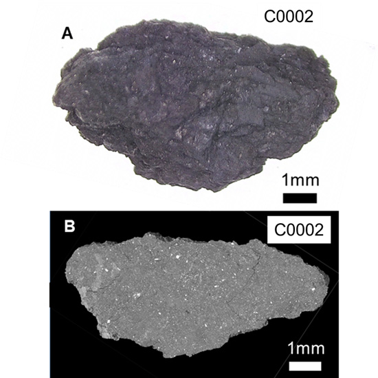
The teams from the IAS and the SMIS beamline of the SOLEIL synchrotron focused on studying the mineral composition of different samples from Ryugu, ranging from small grains of a few microns to millimeter-sized stones. This work used infrared hyperspectral* imaging (2D and 3D) in a wide range of wavelengths coupled with Raman microscopy and electron microscopy (ICMMO, CentraleSupelec), thanks to sample preparations developed specifically for this mission (collaboration with CentraleSupelec and IEMN-Lille). The infrared spectra of Ryugu grains have been compared to those of different primitive meteorites to determine its origin and evolution.
At the SMIS beamline, hyperspectral maps were acquired on sub-millimetre samples of Ryugu asteroid in the mid-infrared range (wavelengths between 3 and 15 µm) at the highest spatial resolution available - measurements every 10 µm approximately. Additional global spectra were recorded for each sample in the far infrared (wavelengths between 15 µm and 1 mm).
These hyperspectral maps reveal the variability of functional chemical groups on the scale of a few micrometres and the intimate association of phyllosilicates and aliphatic components of the asteroid. These measurements show that the Ryugu samples brought back to Earth by the Japanese JAXA Hayabusa 2 mission are broadly similar to CI1-type chondrites (carbonaceous stony chondrites), but with a significantly higher level of CH2/CH3 chemical groups than is generally measured in this type of chondrite collected on Earth.
* Hyperspectral imaging: by coupling absorption microscopy and spectroscopy on a zone of the sample, simultaneously at several wavelengths (in the infrared range, in this case), a map of the chemical composition of the zone studied is obtained.