The behaviour of foie gras during cooking
During the festive season, duck foie gras is a product particularly appreciated by French people. Cooked in verrine before being arranged in (unflattened!) slices, or fried, it is being consumed in multiple ways. However its quality remains uneven in spite of the standardization of duck farming and production processes. Cooking leads to a more or less intense fat loss, which induces a variability of the cooking yield and sensorial qualities. A team from INRA compared the protein matrix of foies gras presenting different levels of fat loss during cooking (from 9 to 33% of fat loss). Their results, published in the Journal of Agricultural and Food Chemistry establish a relationship between the fat loss due to cooking and the composition of the protein matrix of foie gras.
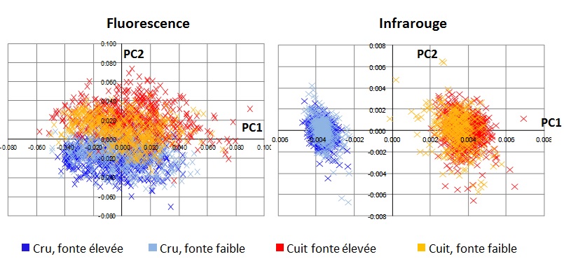
The protein matrix was analyzed in situ on thin sections (10 µm) of foie gras samples, collected before and after cooking and fixed by ultra-fast freezing. The morphology of the protein matrix was characterized by histology after staining the structures. Chemical composition was analyzed by infrared microspectroscopy (SMIS beamline) and UV fluorescence microspectroscopy (DISCO beamline) on a tissue section free of any additive.
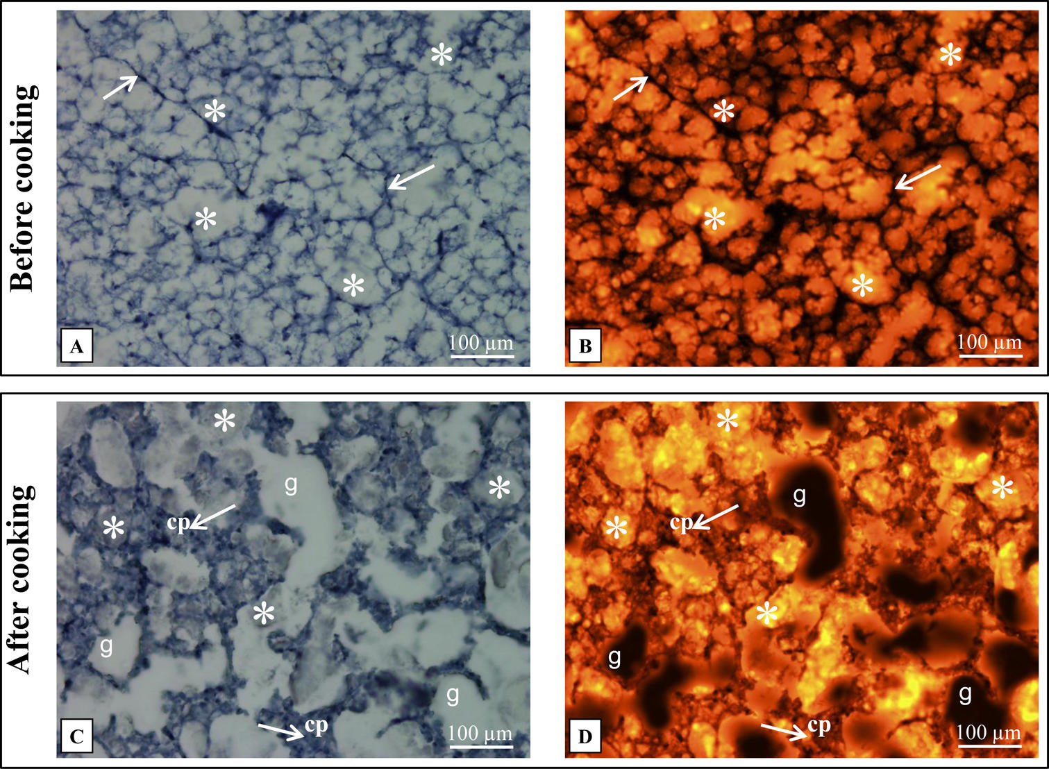
Before cooking, the protein matrix presented a rather thin structure (Figure 1) and the lipid droplets were small. After cooking, the matrix had thickened, due to heat-denaturation and protein aggregation, and the lipid droplets were bigger, suggesting the merging of smaller droplets during heating. These findings are all the more true when the fat loss during heating is important.
Infrared microspectroscopy gave the scientists some information on the molecular structure of proteins, when fluorescence microspectroscopy that characterized the self-induced fluorescence attributed to tyrosine, tryptophan and collagen, gave information on the composition of tissue medium. Statistical analysis of spectra revealed a sensible evolution of composition and molecular structure of proteins during the cooking process. Furthermore, spectral differences depending on the level of fat loss were observed, before cooking as well as after. These effects, however small, are significant enough to consider distinguishing (before cooking) the foies gras that are likely to loss fat during the heating process, and rather use them as process products.
In the end, scientists coupled several analysis techniques that showed that despite the standardization of production and processes, the biophysical characteristics of rough foie gras can vary and that all foies gras do not react in the same way when heated. Thierry Astruc, one of the authors of this study confirms the contribution of the synchrotron: “Chemical imaging and localized spectroscopy techniques allow characterizing the biological materiel in situ while minimizing the modifications linked to sample preparation. Coupling these studies with synchrotron radiation allows us to take advantage of the wavelength continuum and select those which are most relevant to amplify spectral response. Spatial resolution is sensibly better and the beamline scientists’ expertise, concerning equipment as well as data processing, ensures the reliability of the results.
When proteins are less sensitive to heat, the lipidic retention is better and allows a better quality of foie gras. There is no doubt that scientists have been able to taste the nuances of flavor between different samples…