SEXTANTS beamline is dedicated to the study of magnetic and electronics properties of solids by polarized soft x-ray elastic and inelastic scattering. The beamline cover the energy range from 50 - 1800 eV (optimized from 70 to 1200eV)
The purpose of the beamline is first and foremost to deliver the highest flux possible within a small focused beam over the 100 to 1000 eV spectral range, with good performance over an extended range going from 50 to 1700 eV. The flux at the sample position should exceed 1013 photons.s-1 over the optimized energy range.
This will involve two tunable undulators in a medium-length straight section and a monochromator designed to provide a resolving power in excess of 10000 at full throughput in a ~ 20 µm x 80 µm spot at the exit slit. Further, a set of refocusing mirrors (some of them bendable) will be available to focus the beam to three distinct working areas at two branch lines. Switching mirrors will enable the alternate use of the two branch lines. Refocusing optics will be adapted to each experimental technique. A very small vertical spot (~2 µm) will be necessary for high resolution inelastic scattering. A more symmetric spot (80 µm x 50 µm H x V) will provide optimum performance for coherent scattering. Finally, elastic scattering and diffraction experiments can require different conditions. Bendable mirrors will make it possible to adjust the focal distance, spotsize or beam divergence to the specific needs. Many experiments will be dealing with magnetic samples or involving orbital symmetry selection, thus helical undulators providing variable polarization have been specified.
The beamline will serve three types of experiment involving four instruments, three of them already financed through CNRS, ANR and cooperation with outside laboratories.
- Resonant inelastic x-ray scattering (RIXS)
- X-ray resonant magnetic scattering (XRMS), 2 instruments
- Coherent x-ray scattering and Fourier transform holography
These are photon-in-photon-out resonant techniques, highly appropriate to the assessment of element specific magnetic properties under applied magnetic fields, with tunable bulk versus surface sensitivity.

Team


Research associates
- Sorin Chiuzbaian (LCP-MR, Paris VI)
- Jean-Marc Tonnerre (Institut Néel, Grenoble)
- Maurizio Sacchi (INSP Paris VI Umr 7588)
Video
Early September 2018: a user group coming from Frankfurt Goethe-University is unloading a truck, in order to install an experimental set-up on SEXTANTS beamline. This set-up measures the angular distribution of the photoelectrons emitted from a molecule of interest after absorption of a synchrotron radiation pulse. The experiment will last a week. Then, the users will reload the truck and go back to Frankfurt, waiting for their next experiment on SEXTANTS, in 2019.
Technical data
50 – 1700 eV
E/ΔE > 104 up to 1200 eV
Full range: 50 - 1700 eV
Optimized: 70 - 1500 eV
Circular left/right, Linear horizontal/vertical
2×10¹⁵ Phot/s/0.1%bw @ 100eV
1×10¹⁴ (100 eV) - 2×10¹³ (1000 eV) ph./s/0.1% bw (estimates)
M1 A/B : plane/spherical mirrors in horizontal deviation
Monochromator: 5 VLS plane gratings + M2 (spherical mirror)
M3/M6 toroidal switching mirrors on hexapod, feeding two branches, one for inelastic (M3), the other for elastic (M6) scattering.
A. Inelastic scattering branch. Elliptical refocusing mirror.
B. Elastic scattering branch. Intermediate focal point (M6).
C. Elastic scattering branch. Two bendable refocusing mirrors
A. 80×2 μm2
B. 80×50 μm2
C. between 90×15 μm2 and 190x30 μm2 (bendable mirrors)
A. RIXS spectrometer.
B. Coherent scattering (FTH) or resonant scattering (IRMA)
C. Resonant scattering (RESOXS, IRMA) or user equipment
Scientific Opportunities
| Resonant Inelastic Xray Scattering (RIXS) |
|
|---|---|
| Soft X-ray scattering and Resonant Magnetic Scattering (XRMS) |
The Sextants beamline hosts two reflectometers, RESOXS (low-T, high fields, ultra high vacuum) and IRMA (compact to fit in B working area, quick access in high vacuum). |
| Imaging by coherent X-ray scattering and Fourier Transform Holography (FTH) |
FTH imaging of magnetic domains with spatial resolution better than 50nm was achieved during the commissioning of the Sextants beamline.
|
X-ray Excited Optical Luminescence (XEOL) developed at SEXTANTS RIXS endstation
Alessandro Nicolaou, Alberto Zobelli, Victor Porée, Laura Susana, Mathieu Kociak, Jean-Denis Blazit, Victor Pinty
X-ray Excited Optical Luminescence (XEOL) is a technique combining X-ray absorption and optical luminescence to explore the electronic and structural properties of materials. By exciting a sample with X-rays and analyzing the emitted optical photons, XEOL provides insights into local electronic structure, defects, and dopant interactions, making it particularly valuable in materials science. The integration of XEOL with Resonant Inelastic X-ray Scattering (RIXS) at the inelastic branch of the SEXTANTS beamline represents a quite unique experimental setup allowing for high-resolution advanced measurements that can directly correlate with structural and electronic properties of materials.
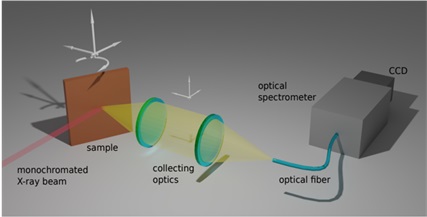
Figure 1: Sketch of the XEOL Setup installed at SEXTANTS.
Our XEOL system, shown in Fig. 1, features two interchangeable lenses (UV fused silica and UV-VIS coated plano-convex) for precise light collection via an optical fiber bundle. This system is connected through an optical fiber to a « Princeton IsoPlane® Advanced » spectrometer coupled with a ProEM® 1600 EMCCD camera, sensitive across the NIR-UV range. It enables measurements in the energy range of 1 to 6 eV (200–1200 nm) with an overall energy resolution down to 1 meV, as shown in Fig. 2. It operates within a temperature range of 18 K to 350 K, with the ability to apply a voltage on the sample and/or a magnetic field up to 0.45 T.
Figure 2 displays XEOL spectra acquired at 18 K for: (a) heavily carbon-doped h-BN, showing a narrow 4.1 eV zero-phonon line followed by complex phonon replicas; (b) pure h-BN, highlighting characteristic strong far-UV excitonic features [1]
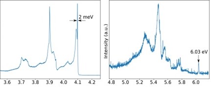
Figure 2: XEOL spectra acquired at 18K for an heavily carbon doped ℎ–BN crystal where it appears a narrow 4.1 eV zero phonon line emission followed by complex phonon replica (left) and for a pure h-BN crystals where the characteristic strong far UV excitonic features can be detected at about 6 eV
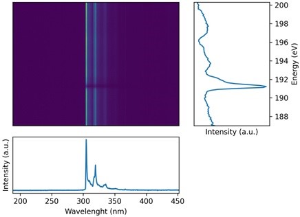
Figure 3: XEOL spectra acquired as a function of the X-ray excitation energy across the B K-edge
The system is coupled with the beamline's automation and acquisition control system, enabling full luminescence spectra acquisition across varying incident X-ray photon energies, as shown for cBN in Fig. 3. Real time post-processing techniques enable precise structural-luminescence correlations, disentangling the different structural phases present in the system.
Finally, the XEOL setup at RIXS branch of the SEXTANTS beamline represents a significant advancement in simultaneous structural and luminescent characterization, opening novel possibilities in both fundamental and applied materials science.
Main Characteristics:
X-ray excitation: 50-1600eV
- Energy Range: 1 to 6 eV (200–1200 nm)
- Energy Resolution (XEOL): 1 meV
- Temperature Range: 20 K to 350 K
- Electric and Magnetic Field: Applied 2D vectorial field (up to 0.45 T)
[1] Bright UV Single Photon Emission at Point Defects in h-BN R. Bourellier et al. ACS Nano 13;16(7):4317-21 201
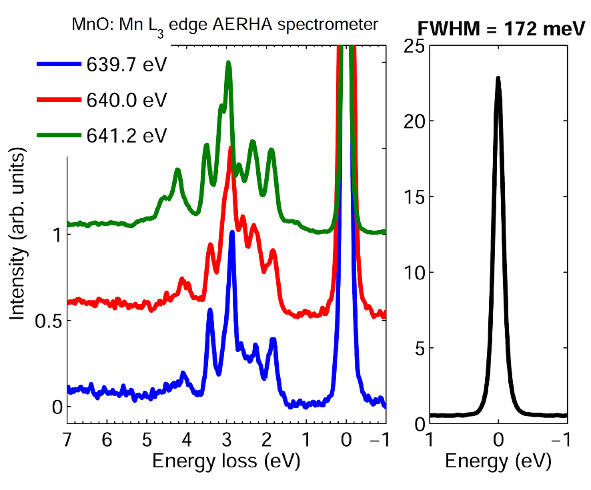
Status of AERHA RIXS Spectrometer April 2016
- Energy range : 50 - 900 eV
- Typical combined (beamline + spectrometer) energy resolution in standard operation (end of 2013)
- Copper M edge 18-20 meV
- Carbon K edge 80-90 meV
- Nitrogen K edge 100-110 meV
- Oxygen K edge 120-130 meV
- Manganese L egde 160-170 meV
* For higher energy resolution please contact the beamline staff
* the tradeoff between spectrometer transmission and resolution can be adapt on demand (x-ray sensitive sample, RIXS map..)
- Collimating mirror undercommissioning (A gain of flux between 2 and 3 has been measured depending the energy range) Planned to be open to user by end of 2016
- Angle resolved RIXS:
* Sample rotation open to user
* Spectrometer rotation: to be commissioned
- Sample temperature:
* 20 -300K open to user
* > 300K : please contact the beamline staff
- X-ray absorption spectroscopy:
* TEY available
* FY available
- New Liquid/gaz cell for RIXS: Developed in collaboration with MAX IV
* gaz : open to user
* liquid: please contact the beamline staff
Dec 2014:COMET Instrument
COMET: a new endstation for COherent Magnetic scattering Experiments in Transmission
This UVH end-station is placed in the intermediate focal point of the elastic branch of the Sextants beam-line. It is specialized in transmission soft x-rays coherent diffraction and particularly coherent imaging, using the Fourier transform holography and ptychography techniques. The chemical selectivity combined with the fully controlled photon polarization makes it a powerful tool for the study of artificial nano-structured magnetic materials.
The sample environment is very flexible, with two independent encoded 3D stages for the mask and the sample. The robust under-vacuum sample holder allows extreme conditions, including low temperature, magnetic coils or RF pumping devices. These blocs are placed on an intermediate rotation stage for fast changing of the transmission angle (+/-45°). Additional piezo rotation stages can be mounted on the mask and sample holders for custom user experiments.
Internal setup: sample environment
Internal setup: CCD
The 2D detector is a back illuminated, 2048x2048 CCD, with 13.5 µm pixel size. The sample to CCD distance can vary from 5 to 45 cm. An XZ piezo controlled beam-stop is integrated to the CCD head. An Al 100 nm filter is protecting the cip from the visible light.
This instrument was developed by the Sextants beam-line team (H. Popescu, N. Jaouen, M. Sacchi (associate), R. Gaudemer) in collaboration with the LCPMR team (J. Perron, R. Delaunay, R. Vacheresse, B. Pilette and J.Luning). The holography masks and coherence pinholes were FIB fabricated at CSNSM by F. Fortuna.
Technical specifications:
- UHV environment
- High mechanical stability
- Low temperature: 130K
- Magnetic field: 700 Oe in plane (V and H) in pulsed mode/50 Oe in static
- Mask stage: XYZ, 21 mm travel, 500 nm encoded resolution
- Sample stage: XYZ, 35 mm travel, 20 nm encoded resolution
- motorized rear diode (AXUV100, Al capped)
- intermediate rotation table for turning together the mask and sample blocs (+/- 45°)
- 2D detector: 2048x2048 back illuminated 16 bit CCD detector, distance to sample
from 5 to 45 cm
- Integrated beam-stop (2 available sizes) motorized under vacuum in XZ directions, with 50 nm encoded resolution
- beam attenuators in the upstream part of the elastic branch: from 20 to 1000 nm gold films
- piezo-shutter driven by the CCD controller
All these equipments are controlled through the Soleil tango-based software system and automatic data acquisition is possible via the ipython environment. This station is now opened to the user community.
Station manager: Dr. Horia Popescu
Beamline manager: Dr. Nicolas Jaouen
Nov 2013: Status of AERHA RIXS Spectrometer
from: Chiuzbaian et al;Review of Scientific Instruments, 2014, 85(4): art.n° 043108
- Energy range : 50 - 900 eV
- Typical combined (beamline + spectrometer) energy resolution in standard operation (end of 2013)
Copper M edge 18-20 meV
Carbon K edge 80-90 meV
Oxygen K edge 120-130 meV
Manganese L egde 160-170 meV
* For higher energy resolution please contact the beamline staff
* the tradeoff between spectrometer transmission and resolution can be adapt on demand (x-ray sensitive sample, RIXS map..)
- Collimating mirror undercommissioning (A gain of flux between 2 and 3 has been measured depending the energy range) Planned to be open to user in 2014
- Angle resolved RIXS:
* Sample rotation open to user
* Spectrometer rotation: to be commissioned
- Sample temperature:
* 24 -300K open to user
* <24 K under commissioning
* > 300K : to be commissioned
- X-ray absorption spectroscopy:
* TEY available
* FY is under development: open to user in 2014
- New Liquid/gaz cell for RIXS:
Under development in collaboration with MAX IV
First experiment schedule in first semester 2014
Open to external user in second half of 2014
April 2011: Measured Flux at the sample position for the RIXS spectrometer

2011: Commissioning experience CXI-FTH
First tests of the Fourier Transform Holography set-up on the SEXTANTS beamline started in October 2010, using a temporary endstation (collaboration with J. Lüning, LCP-MR). Samples were Co/Pd multilayers fabricated by R. Delaunay (LCP-MR, Paris VI), using magnetron sputtering. Nanostructuring of the samples was carried out by F. Fortuna (CSNSM, Orsay), using Focused Ion Beam (FIB) etching. FIB is used to define the area to be imaged (1.6μm in our case) and the reference holes for the holography, whose size enter in the definition of the ultimate spatial resolution.
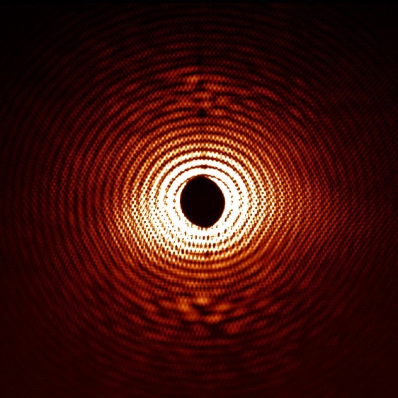
Fig. 1 shows the coherent scattering diagram collected using an in-vacuum CCD. The image size corresponds to 190μm-1 in reciprocal space. Data were collected at the Co-L3 edge energy (780eV), using circularly polarized X-rays. Co/Pd multilayers are homogeneous, apart from their magnetic structure. Worm-like domains with perpendicular up/down magnetization form in the Co layers, originating regular structures with sub-micron period.
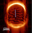
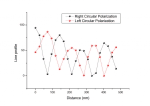
Fogure 2 compares the Fourier transform of coherent scattering patterns obtained with opposite helicities of the photons. Both images display light/dark modulations, related to the magnetic structure of the sample. Line profiles show clearly that these modulations are exactly out of phase when the light polarization is inverted, confirming their magnetic origin. Therefore, we can highlight pure magnetic contributions to the coherent scattering by image subtraction, eliminating all other structural or spurious effects.
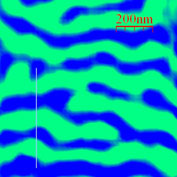
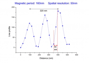
Fig. 3 shows the result of this subtraction: magnetic domains with a period of the order of 150nm are imaged with an overall spatial resolution better than 50nm.
June 2010: First beamline commissioning
Beamline commissioning started in June 2010. Focusing and stability of the M1A/B mirrors under full undulator power conditions have been tested. Absorption measurements with monochromatic X-rays (C-06 cell of the beamline) have been used to characterize the energy/angle curve of the four gratings already installed (G1-G4), covering the 50-1500eV range. Also, the VGD (variable groove depth) behaviour has been addressed. VGD gratings feature a groove depth that depends on the lateral position. Since the photon beam has a very small lateral size at the grating (about 200 microns), by adjusting the lateral position of the grating one can choose a precise value for the groove depth, with different efficiencies for first and second order scattering. Therefore, one can maximize either throughput or spectral purity.
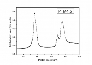
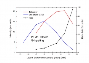
The two figures above show an exemple of the VGD function at the Pr-M5 absorption edge, using G4. By a lateral displacement of the grating over 20mm, one can either minimize 2nd order contamination, which varies by a factor of 20, or maximize 1st order throughput
Resonant Inelastic X-ray Scattering
Sorin Chiuzbaian, Coryn Hague, Renaud Delaunay, Jan Lüning and Stefania Brignolo, LCP-MR, University P. and M. Curie, Paris
Nicolas Jaouen, Cédric Baumier and Maurizio Sacchi, SOLEIL
RIXS, also called resonant x-ray Raman scattering, just like XAS, uses a monochromatic photon beam to promote an electron from an inner shell to an unoccupied outer level. But, contrary to XAS where it is usual to record the total electron or fluorescence yields, the experiment consists in a detailed analysis of a specific radiative decay channel. In a Raman process, the energy of the scattered photon ω is less than the excitation energy Ω; it is resonant when Ω corresponds to the exact difference between two excited states. Because radiative decay in the soft x-ray region is orders of magnitude smaller than non-radiative decay and because spectra are recorded within a narrow band of energies, only third generation synchrotron radiation sources provide the flux and brightness needed for these experiments. In recent years, important new results have activated new theoretical studies which show that a whole new field of spectroscopy is opening up which justifies investing in this type of research at SOLEIL.
- The study of X-ray absorption edges with improved resolution no longer limited by the lifetime of the intermediate state.
- Observation of low-lying excitations associated with the ground state. The information obtained is analogous to that obtained from optical spectroscopy except that the core-hole excitation makes it an element selective technique.
- Band mapping. High-resolution in excitation and analysis is needed. Future developments should make it possible to perform angle resolved RIXS. The potential advantage over ARUPS is, here again, element selectivity and bulk sensitivity.
- The use of polarized radiation underlines symmetry effects and especially magnetic properties. Measurements may be performed in the presence of an applied magnetic field. RIXS is highly responsive to the degree of localization of the core-hole excited intermediate state, itself an important parameter in our understanding of magnetic properties.
- The bulk sensitivity of the RIXS technique may be fully exploited to study buried layers. Examples of such experiments would typically be concerned with the study of buried ultra-thin oxide layers such as MgO, NiO, or V2O3.
- Lastly, RIXS should serve the fundamental interests of theory and computational methods applied to core-hole spectroscopies such as resonant Auger spectroscopies. Testing theories will certainly benefit from being able to compare both types of radiative and non-radiative experiment and improved conditions for gas phase studies. High flux and brightness are obviously indispensable to experiments involving low density samples.
A high resolution grating spectrometer was designed in 2002. It is based on a plane ruled-grating with variable line-spacing and a spherical mirror. A large additional mirror to improve the collected angle in the horizontal plane will optimize efficiency and the signal-to-noise ratio. A high-efficiency 1024x1024 13µm pixel back-illuminated CCD detector was used in a fixed position. In principle, the ultimate resolving power of such a design is over 10000. In practice, the resolving power of the instrument was around 1000 for a 5 µm (vertical) source size. The spectrometer is of a compact lightweight construction (total length 1.5m) and has been fully tested at various synchrotron sites.
Based on this experience, the project of a new high resolution spectrometer has been funded by the French ANR. The instrument will have a similar optical design as the prototype, with an elliptical cylinder replacing the spherical mirror and an overall size of 3.5m. Coupled to a small vertical spotsize of the incoming x-rays (90% of the intensity in less than 2 microns), the new spectrometer will have an improved resolving power of up to 8000 and always in excess of 5000 over the energy range 50-1000 eV.
X-ray Resonant Magnetic Scattering
Nicolas Jaouen and Maurizio Sacchi, SOLEIL
Jean-Marc Tonnerre, Stéphane Grenier and Marta Elzo
(Institut Néel - CNRS, Grenoble)
Though de Bergevin and Brunel were the first to demonstrate magnetic X-ray scattering as early as in 1972 using a conventional X-ray source, it was not until the late 1980's that very large X-ray magneto-optical effects were predicted and demonstrated experimentally. This breakthrough was due to the possibility of tuning the energy of the photon beam to the onset of a core absorption edge of a magnetic element.
The first X-ray resonant magnetic scattering (XRMS) experiments were performed in the 4-10 keV energy region, next, even larger magnetic effects at core resonances located in the soft X-ray region (50-2000 eV) were observed in the 3d transition metals (TM) and rare-earths (RE). This is explained by the direct involvement of the magnetic orbitals in the scattering process (3p,2p to 3d resonances in TM's and 4d,3d to 4f in RE's). Soft X-ray experiments have since been performed both in the specular reflectivity mode and the Bragg diffraction mode on multilayers.
The specific properties of soft X-ray resonant scattering that make it a major tool for investigating electronic structure and magnetism may be summarized as follows:
- Inherent sensitivity to structural properties.
- Element selectivity and sensitivity to electronic properties.
- Sensitivity to magnetic properties and magnetic structure (magnetization depth profile, roughness, domain size, etc.)
- Large magneto-optical effects, due to direct involvement of magnetic orbitals in the electronic transitions. The high sensitivity makes it easy to investigate surface layers (down to a single atomic layer). The tunable photon penetration depth permits the analysis of buried thin layers
- Flexibility in the choice of experimental geometries: ferromagnetic (FM) and antiferromagnetic (AF) ordering, probed by circular or linear polarization
- Imperviousness to strong or time dependent magnetic fields and to charging effects (photon-in-photon-out experiment).
Supported by adequate computational models, the large experimental input provides what is often a unique path to information concerning the electronic and magnetic properties of materials, in particular where the interplay between structural and magnetic properties is of interest.
In terms of new perspectives, two directions seem very promising, namely off-specular magnetic scattering and time-resolved analysis. The former has been the subject of investigation over the last few years, and has been used recently to address interesting problems like magnetic roughness and order in closure domains. The latter is more at an exploratory stage, but first results indicate that time resolved experiments would give new opportunities to investigate magnetization dynamics with all the advantages of resonant magnetic scattering of polarized soft X-rays.
New directions in resonant scattering (e.g., diffuse and time resolved scattering) are definitely demanding a high flux. To stick to what is already known, the analysis of specular and diffuse (magnetic) scattering over wide q ranges means spanning at least seven or eight decades in intensity. Therefore, 1011 ph.s-1 should be considered an absolute minimum flux for future XRMS experiments. Including dynamics in our objectives can only push this limit upward.
As far as the spot size is concerned, the requirement of a small beam divergence is difficult to combine with a small beam. We have found a satisfactory compromise by relaxing focusing conditions for the XRMS endstations. A small spot is nonetheless necessary, since magnetic studies are more and more oriented towards small objects As an example, dynamic studies are better performed on small samples directly prepared on micromagnets, with typical lateral size of a few tens of microns. We believe that a beamline that can deliver an intense flux (1013 ph.s-1) in a small spot of variable size (from 10 µm x 50 µm, vert. x horiz., up to 40 µm x 200 µm) will open up new opportunities in the field of soft X-ray resonant magnetic scattering.
Two groups in France are strongly involved in the development of these techniques and have undertaken the construction of two experimental set-ups with complementary concepts.
Instrument 1. M. Sacchi and co-workers have developed at LURE a new instrument for soft X-ray scattering, that is intended to be part of the experimental set-up of the beamline. The instrument (IRMA: Instrument pour la Réflectivité MAgnétique) meets the criteria for surface science experiments, including in situ preparation and characterization in a UHV environment (10-10 mbar base pressure). This is a strict requirement for extending present resonant scattering studies to the domain of surface science. Though the high sensitivity of soft X-ray scattering permits the investigation of very thin surface layers, up to now, these kind of studies have been held back by the absence of reflectometers working under UHV conditions. IRMA is now based at the synchrotron source ELETTRA (Trieste, Italy), where it is used by the french community as well as by international users.
Instrument 2. J.-M. Tonnerre and co-workers at the Laboratoire de Cristallographie (now Institut Néel) in Grenoble have implemented a large volume reflectometer under high vacuum (10-8 - 10-9 mbar). The availability of such a set-up (named RESOXS) permits to investigate, statistically, the magnetic morphology of nano-particles. These may be either self-organized, buried, elaborated on patterned substrates, or prepared by microlithography techniques. It is possible also to probe the collective behavior of an assembly of nano-particles with respect to an external applied magnetic field or to a temperature change.
The experimental set-up includes a quadrupole electromagnet, designed to apply an in-plane magnetic field in any direction, and sample holders designed for low or high temperature measurements.
An extension of the project (in collaboration with SLS) concerns the development of a polarization analyzer mounted on the detector arm. The RESOXS reflectometer was installed at the Swiss synchrotron source SLS and is now installed at SOLEIL.
Lensless X-ray Microscopy via Scattering of Coherent X-rays
Horia Popescu, Nicolas Jaouen and Maurizio Sacchi, SOLEIL
Jan Lüning, LCP-MR, University P. and M. Curie, Paris
A revision of the original beamline design made available an extra experimental location, at the intermediate focal point of the XRMS branch, that was not foreseen in the original beamline proposal and was not included in the original funding plan. The recent demonstration of high-resolution lensless microscopy by scattering of coherent x-ray beams has motivated us to propose the development of this novel technique at the SEXTANTS beamline, a project that was presented to the SOLEIL Scientific Advisory Committee in November 2006.
Apart from the appealing novelty and elegance of lensless microscopy, the main arguments for the world-wide interest in this technique are its potential for higher spatial resolution as well as for its compatibility with extreme sample environments. The scientific motivation for the proposed development is based on our interest in magnetization dynamics occurring at the nanometer length scale. These experiments would become feasible at SOLEIL by combining the high spatial resolution of the instrument with the intrinsic time resolution of SOLEIL's picosecond short x-ray pulses.
Our goal is to develop a versatile instrument for novel x-ray microscopy techniques based on coherent scattering serving both transmission and reflection geometries. Techniques the instrument shall enable include Fourier Transform holography [2-4], phase retrieval microscopy, ptychography and lensless microscopy with curved wave fronts. Seed funding for this project has been requested by a proposal to the PNANO program of the ANR, in collaboration with LCP-MR (Paris VI) and CSNSM (Paris XI). The proposed instrument for scattering based imaging would immediately enable us to overcome today’s main shortcoming of x-ray microscopy, namely, the achievable spatial resolution. While about 50 nm, which can routinely be provided today for scientific applications, is an outstanding value for a wide variety of applications, there is an increasing demand for sub 10 nm spatial resolution, driven especially by the growing importance of nanoscience and nanotechnology. The common principle of scattering based microscopy is to replace the resolution limiting optical element with a two-dimensional detector and thus to record the information contained in the scattered electromagnetic wave front without introducing any distortions. In the absence of wave front degradations, sub 10 nm spatial resolution can be realized, since x-ray wave lengths measure only a few nanometers or less. The key properties setting the proposed instrument apart from other microscopes are:
- potential sub 10 nm spatial resolution. In lensless microscopy by scattering of coherent x-rays the achievable spatial resolution is only limited by the wavelength, which is a few nanometer or less for the x-ray photon energy range.
- versatile in situ sample manipulation. The absence of any optical element next to the sample facilitates the installation of the equipment necessary to manipulate the sample in situ, for instance for applying strong fields, attaining extreme temperatures or working in a controlled atmosphere.
- flexible sample environment. Conventional, lens-based x-ray microscopes are seldom compatible with ultra-high vacuum (UHV) due to the necessity of in-vacuum motors and controls for optics-sample alignment. Our instrument, being truly UHV compatible, will fill this gap. At the same time, various sample environments ranging from ambient pressure to the presence of liquids can be implemented by making use of the available space for appropriate sample enclosures.
- insensitivity to vibrations and thermal drift. The technique is inherently insensitive to vibrations and thermal drifts, since real space resolution is achieved by detecting the scattered intensity in Fourier space, the angles being resolved by micron sized detector pixels. Nanometer spatial resolution can be achieved with micrometer mechanical stability, which can be obtained without specific means of vibration damping or thermal isolation. This will result in a very compact setup that may be used eventually at different beamlines of SOLEIL.
- extended energy range. There is no fundamental limitation for the photon energy range at which this instrument can be used. Within the energy range of the SEXTANTS beamline one finds in particular the K edges of the light elements, relevant for studies on organic materials; the L edges of transition metals relevant for studies on charge and spin ordering phenomena in transition metals and their compounds, as well as the M-edges of the lanthanides.