From yellow to white and back again: the secret of crab spiders’ color changes
Animal coloration keeps fascinating us. While the process of pigmentation is well understood in most animals (including humans), the process of depigmentation is much less so. By studying color changes of spiders, researchers have begun to unlock the secret of pigment degradation.
The mechanisms that allow cells to degrade pigments, or even to recycle them, are necessary in contexts such as vision, when the pigmented cells of the retina are confronted with a large flow of light that can damage them, or during color changes in some animal species. This is the case of some crab spiders, which can reversibly change from white to yellow depending on the flower on which they hunt; a form of camouflage that could give them an advantage in capturing preys.
By studying these pigmentation and depigmentation mechanisms with a combination of advanced imaging techniques, scientists from the University of Tours, the CNRS and the Curie Institute, in collaboration with researchers from the NANOSCOPIUM beamline and the CryoCapCell company, have discovered that crab spiders’ pigment organelles (intracellular "factories and storage sites" for pigments) are similar to pigment organelles of humans, snakes and insects, notably because they all derive from the same precursor and naturally accumulate metals.
The NANOSCOPIUM beamline has played a key role in detecting these metals within sub-micrometer sized organelles at different stages of pigmentation. By coupling the high sensitivity of the X-ray fluorescence technique to a unique multi-lengthscale scanning system implemented on the beamline, it was possible to identify zinc, calcium or cobalt at micro- and nanoscopic scales, in complex and variously colored biological samples.
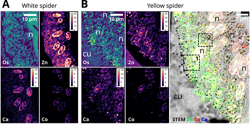
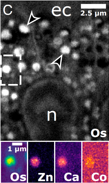
The results obtained allow to classify pigment organelles of crab spider in the large family of lysosome-related organelles. Lysosomes are one of the main actors of cellular content degradation, indicating that pigment organelles also possess such capacity and thus have the ability to degrade their pigments. This hypothesis is confirmed by the 3D description of pigment organelles whose contents were undergoing degradation in bleaching spiders.
Beyond the case of crab spiders, these results would indicate that all pigment organelles of animals could possess the same degradation faculties, and that the mechanisms involved in color changes could also function in other contexts: crab spiders open new research perspectives to better understand the life cycle of pigments.
This study model and these results are to date the most precise on the recycling phenomena of pigments supposed to exist in the human eye.
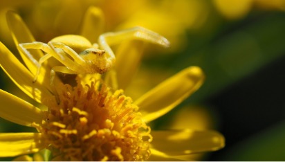
Presentation picture: Thomise variable (Misumena vatia) jaune à l’affut sur une fleur de la même couleur. Crédit : Florent Figon/Flickr – CC BY SA