X-ray nanotomography now available at SOLEIL!
The new X-ray nanotomography instrument on the ANATOMIX beamline at SOLEIL has seen its first official users. The DYSCOL unit (Dynamics and Structure of Liquid Bodies) of the French National Institute of Agricultural Research (INRA) used the instrument to obtain 3D volume images of seeds of an oil- and protein-rich plant on length scales characteristic of intracellular lipid-storage organelles, without physically cutting the specimens or applying staining protocols to enhance contrast. This nano-imaging station has been specifically developed and built by SOLEIL in the framework of the NanoimagesX project funded by the Equipments of Excellence program (EQUIPEX) of the Investments for the Future scheme of the French State.
Recently built at SOLEIL, the beamline ANATOMIX (Advanced Nanotomography and Imaging with Coherent X-Rays) is dedicated to three-dimensional imaging with hard X rays at microscopic length scales. It is one of two “long” beamlines at SOLEIL, which allows its users to benefit from a very coherent X-ray beam and thus obtain images of exceptionally good contrast and resolution.
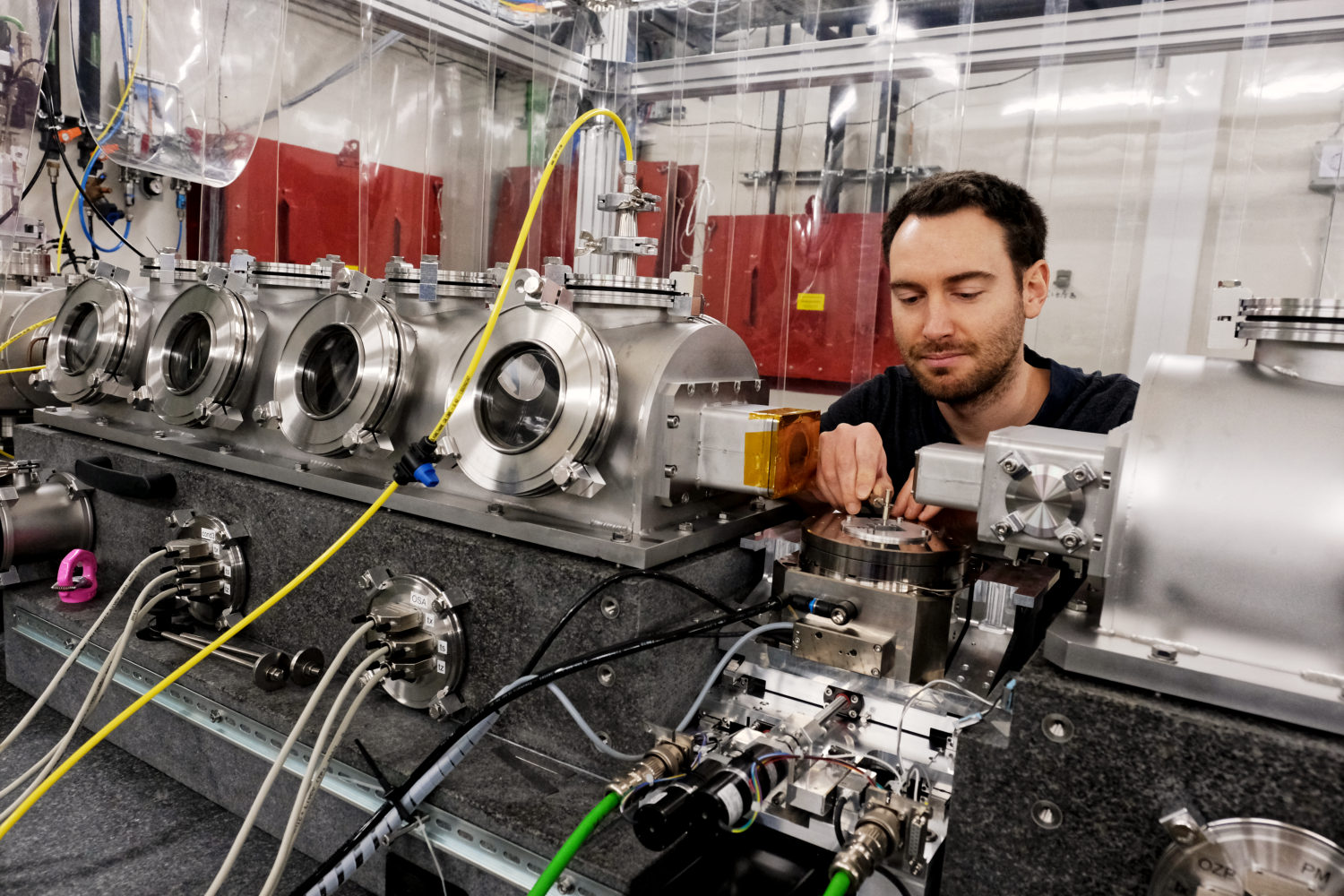
The new nanotomography end station on ANATOMIX (figure a) works in a photon energy range from 5 to 20 keV.
Based on the concept of a full-field transmission X-ray microscope (TXM), it yields a spatial resolution in 3D on the order of 100 nm and below. In complement to X-ray microtomography, which gives access to length scales down to around 500 nm, users at ANATOMIX can now explore complex biological structures or materials systems in three dimensions with a resolution down to 20 nm (pixel size). The Arabidopsis thaliana seeds studied by the INRA team have a diameter around 0.3 mm and were measured without cutting the specimen. The data acquisition time for a full tomographic volume was 2.5 minutes. Figure b shows virtual cuts extracted from 3D volume images obtained with tomography at different length scales up to the full grain.
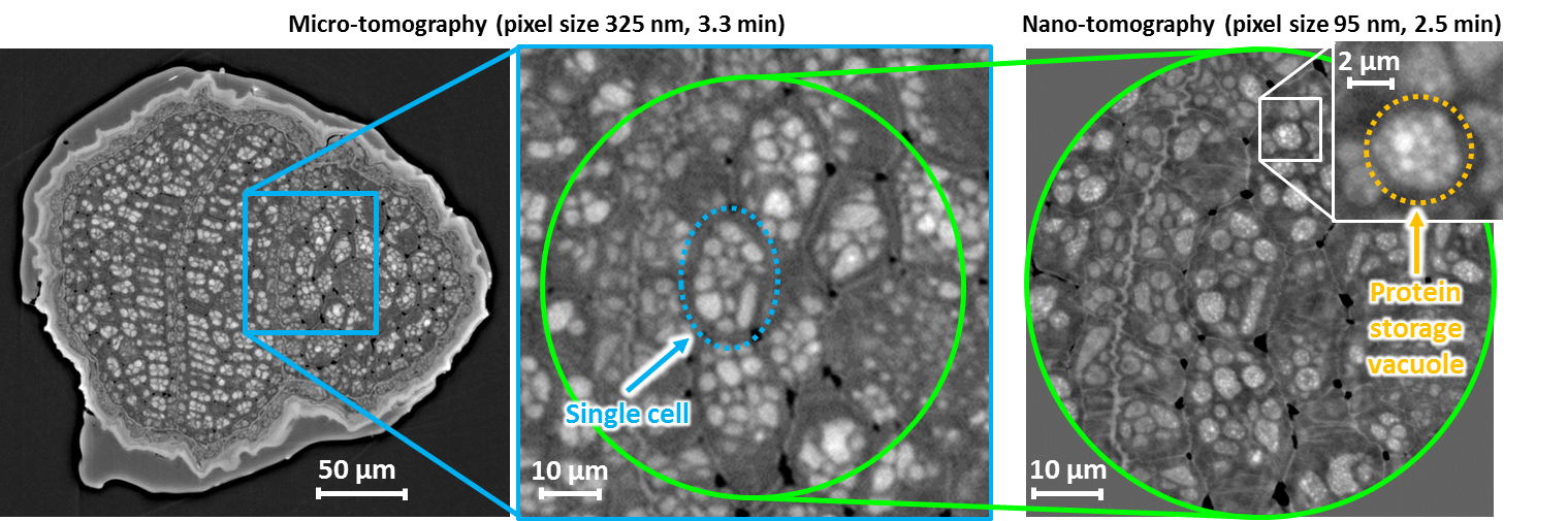
During the four days of the experiment, the user team acquired more than 200 nanotomography and high-resolution microtomography volume scans. For ANATOMIX, it was also the first time that both techniques were combined in a single experiment (figure b).
This first team of external users will soon be followed by several more. If you are interested in submitting a proposal for the next call for projects, contact the beamline scientists!