When paper gets a sun burn at SOLEIL
Of the many physico-chemical analysis and imaging techniques that enable a better understanding, insight or assessment of the state of preservation of cultural heritage objects, the techniques that use X-rays produced by synchrotron radiation (SR-X) are at the forefront. This is due in particular to the nature of the beam, extremely bright, tunable over a wide energy range, stable and providing spectral images with spatial resolutions of up to tens of nanometers while covering fields of view to up to a few centimetres. These beam properties allow probing materials from the perspective of their crystalline structure or chemical composition and environment with a very high detection sensitivity. The drawback is the risk of physically and chemically modifying the materials analysed. This risk, which is still poorly described, constitutes a significant issue in the study of cultural heritage and ancient materials by SR-X, to which the PUMA beamline at Synchrotron SOLEIL is dedicated.
A team of researchers from the Centre de recherche sur la conservation des collections (CRC-MNHN-CNRS 3224), the IPANEMA laboratory (CNRS- MiC-UVSQ USR 3461) and the PUMA beamline at Synchrotron SOLEIL studied the short- and long-term effects on paper of SR-X radiation exposure.
The study focused on the chemical and structural changes that occur in paper during SR-X analysis and investigated the impact of ambient humidity conditions during and after analysis. Is it possible to define "threshold doses" from which chemical or physical changes can be observed? Do these threshold doses depend on the analytical techniques used to characterize the changes? What are the post-irradiation kinetics of the physico-chemical evolution? Moreover, since cellulosic cultural objects are most often preserved under stable thermo-hygrometric conditions, should these conditions be the same during analysis? These are all questions that were addressed by the team at the Centre de recherche sur la conservation des collections, the IPANEMA laboratory and the PUMA beamline at Synchrotron SOLEIL.
In an attempt to resolve these issues, scientists established a methodology based on the irradiation of a model paper, composed of cotton linters, i.e. more than 99% cellulose (Whatman no. 1). Even though the irradiation could have been performed with more accessible laboratory sources, it was decided to perform the experiments at Synchrotron SOLEIL for three reasons: to match as closely as possible the irradiation conditions to the synchrotron experiments, to have a more precise control of the beam parameters and, finally, to deposit a high dose in the sample in a relatively short time thanks to SOLEIL's high flux. The investigations were carried out on the recently opened PUMA beamline, optimized for Heritage Science research. The irradiation was conducted at room temperature, at 7.22KeV, at varying doses and under 3 hygrometric conditions.
The most direct indicators of cellulose degradation are depolymerization and oxidation. Together these reactions lead to a shortening of the polymer chains, and ultimately to a mechanical weakening of the fibres. These chemical "events" lead to other physical manifestations, such as the appearance of UV luminescence and yellowing. These post-irradiation modifications of the paper were monitored using separation and spectro-imaging techniques, which made it possible to conduct the investigation on a broad spatial scale, nanometric (molecular) and microscopic. On the nanometric scale, the depolymerization of cellulose was assessed by size-exclusion chromatography (SEC-MALS-DRI) and the free radicals formed (especially hydroxyl), indicators of auto-oxidation phenomena, were quantified by high-performance liquid chromatography (HPLC-FLD-DAD). On a microscopic scale, UV luminescence and yellowing were recorded by spectrometry and photography. A means of determining the doses absorbed by the paper was set up in order to define which ones induced changes, with particular attention paid to determining the Lowest Observed Adverse Effect Dose (LOAED). The physico-chemical properties of the paper were monitored over a period of two years.
The study found that the samples were impacted at the nanometric scale immediately after irradiation, starting at the relatively low dose of 21 Gy (equivalent to an irradiation time of 30 s). The higher the dose of SR-X radiation absorbed by the paper, the greater the depolymerization and the higher the amount of free radicals. The onset of UV luminescence and yellowing was delayed and staggered over time (figure 1).
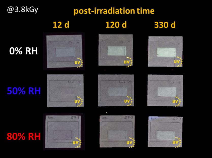
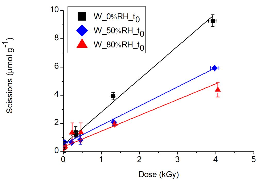
A variation in behaviour under the beam in relation to the paper moisture content was established. The most significant changes in UV luminescence (Figure 1) and depolymerization (Figure 2) were measured for the least humid paper samples, i.e. those irradiated at 0% relative humidity (RH). One of the findings of the research was therefore that it is important to ensure that the paper does not dry out during SR-X analysis.
It was also demonstrated that not all indicators were equally sensitive in detecting changes within the paper. As previously mentioned, depolymerization and the production of hydroxyl radicals occurred immediately after irradiation and over the entire range of doses (7Gy-4 kGy) and RH tested. The fact, on the one hand, that the techniques associated with the measurement of these two molecular indicators are sensitive techniques, and on the other hand, that these changes precede all others, explains why they show the lowest LOAEDs, of 21 Gy for HO° radicals and 210 Gy for glycosidic scissions. These indicators are based on destructive, sample-consuming analyses, and as a result are difficult to implement. Luminescence and yellowing, on the other hand, can be analysed by non-invasive means, and could be implemented for in-situ detection of these paper changes during exposure to the SR-X beam. However, these microscopic indicators appear with a delay of 12 days to 1 year and more (Figure 1), depending on the dose and ambient relative humidity at the time of SR-X irradiation, which limits their usefulness in signalling early physico-chemical changes.
This research has provided information on the potential damage and early detection of SR-X induced changes to cellulosic materials when studied using the PUMA beamline, dedicated to the study of heritage objects. Other questions are currently under investigation, such as the impact of the presence of paper additives (sizing, inks, fillers) and the state of preservation of the object at the time of the SR-X analysis. The follow-up study aims at investigating these aspects and examining the behaviour of ancient cellulosic documents during SR-X beam analysis.