Unraveling the otolith through X-ray nanofluorescence at the NANOSCOPIUM beamline
Otoliths are mineral structures found in the inner ear of vertebrates. In teleost* fish, they continuously form throughout the animal's life, preserving a chronological record of the chemical and biological signatures of events and living conditions over its growth. A valuable indicator of teleost fish lifestyle and environmental variations, otoliths can be challenging to study due to e.g. their small size. Utilizing the NANOSCOPIUM beamline at SOLEIL, nanoscale resolution analysis methods sensitive to the chemical elements constituting the biomineral have allowed the deduction of the environment (river-ocean) and age of pipefish (Teleostei, Syngnathidae) by studying their otoliths at the sub-micrometer scale.
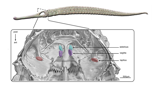
Otoliths, or "ear stones" in Greek, are calcareous concretions in the inner ear of vertebrates (Fig. 1), contributing to stato-acoustic functions (hearing and balance) in the organism. In teleost fish, otoliths serve as valuable biogeochemical information sources, akin to a biological 'black box' due to their continuous growth without resorption, providing a sequential life history of these organisms.
The growth occurs through alternating mineral-rich and organic-rich layers, known as increments. Like tree rings indicating an organism's age, otolith increments allow for age estimations and the duration of significant events. Moreover, during growth, otoliths incorporate chemical elements from the surrounding environment. Microchemical analysis of otolith composition reveals growth dynamics and environmental history, enabling the reconstruction of individuals' migratory and environmental history.
Challenges include detection limits for otolith components, especially trace elements (quantities on the order of parts per million, or ppm), and the resolution of analysis tools, particularly for very small otoliths.
X-ray nanofluorescence spectroscopy (nanoXRF) at the synchrotron can be used to map different elements of interest for otolith biogeochemical microanalysis at the nanoscale. A research program led by the 'Biodiversity Plasticity Adaptation and Conservation' team (UMR BOREA) at the National Museum of Natural History in collaboration with the SOLEIL NANOSCOPIUM beamline achieved the global elemental mapping of otoliths for two species of freshwater pipefish, Microphis brachyurus and Microphis nicoleae (Fig. 2). The spatial resolution was 500 nm for the range of chemical elements detectable by the beamline, from atomic number 13 (Aluminum) to 38 (Strontium).
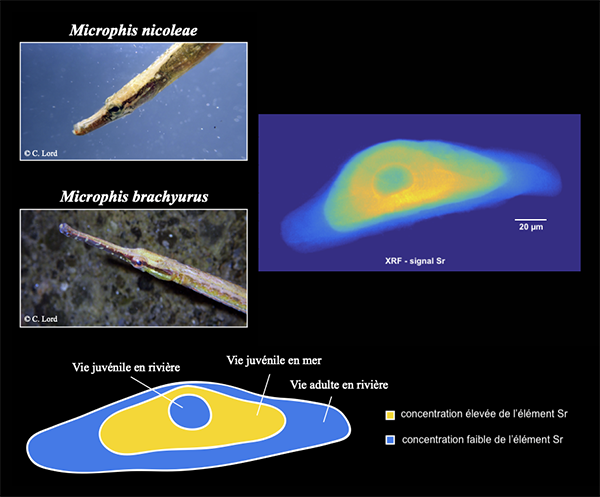
Zonal distribution study of strontium, a chemical element significantly more abundant in marine environments than freshwater, highlighted the signature of an environmental migratory transition. Both studied freshwater pipefish species exhibit a diadromous life cycle (two reverse phases of river-ocean transition). Variation in the distribution of certain trace elements in the otolith (Al, Cu, Fe) may reflect differences in population dispersion routes.
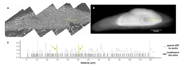
The analysis of the spatial distribution of sulfur has enabled the correlation of its abundance variation with the increment lines on the otolith, thereby providing the opportunity to model a chemical counting of growth rings (Fig. 3). Widely employed in population ecology studies, age estimations were previously conducted through visual counting of growth marks on the otolith, a method limited by the intrinsic visibility of the sample (especially those of reduced size) and the experience of the observer. This innovative sulfur microchemistry counting method, contingent on the need for a high spatial performance detection mode, facilitated age reading on the otoliths of the two studied species, which were otherwise inaccessible to this approach.
The study focused on two pipefish species inhabiting environments vulnerable to anthropogenic pressures, impacting crucial habitats for their life cycle. In the context of biodiversity erosion, this knowledge represents an essential preliminary step for the preservation and conservation of populations.
*Teleosts are a class of bony fishes characterized by the presence of a developed jaw, paired fins, and thin scales.