SIRIUS - Origin of the intense luminescence of two-dimensional supramolecular crystals revealed by GIXD
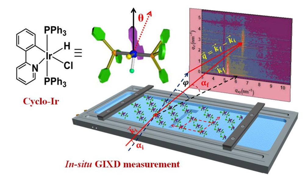
Luminescent materials that continue to emit light after being illuminated find applications in our daily life, especially in lighting devices. For this type of application, two-dimensional (2D) organometallic crystals are very promising. Their luminescence can be strongly enhanced by the phenomenon of aggregation-induced emission (AIE). Understanding and controlling AIE could lead to low-cost photoluminescent 2D materials at room temperature. At SIRIUS beamline, grazing incidence X-ray diffraction (GIXD) technique at the water surface can determine the structure of these crystals.
Two-dimensional (2D) molecular crystals are an important class of materials for applications in electronics and photonics. Their extremely small thickness - ideally a single layer of molecules - explains the name "2D" and gives them unique optoelectronic properties, different from those of bulk organic crystals, while saving material, allowing lower production costs and mechanical flexibility.
Strong luminescence has recently been demonstrated in bulk molecular crystals, exhibiting so-called aggregation-induced emission (AIE) phenomenon. The assembly of molecules causes the restriction of intramolecular rotations and vibrations that could quench luminescence. But the mechanisms involved in AIE in bulk materials can only occur in 2D crystals at very low temperatures (77K, liquid nitrogen temperature) leading to low luminescence at room temperature. Understanding and controlling the self-assembly process of the molecules constituting 2D crystals could allow to obtain low-cost luminescent materials at room temperature.
A team of scientists from laboratories of the Indian Association for the Cultivation of Science (Kolkata) and Saha Institute of Nuclear Physics (Kolkata) have developed a technique to control the fabrication of AIE-active two-dimensional crystals made of cyclometalated Ir(III) complex, [Ir(ppy)(PPh3)2(H)(Cl)] (Cyclo-Ir). The fabrication takes place on the surface of water by the Langmuir monolayer technique, which allows a fine control of the surface density; the stable 2D crystals obtained are of molecular thickness, of a length of the order of µm, and show a luminescence superior to the crystals obtained in solution.
The team came on the SIRIUS beamline to study this system directly on the water surface in order to understand the origin of the luminescence.
Grazing incidence X-ray diffraction (GIXD; see figures) measurements carried out on SIRIUS revealed the formation by self-assembly on the water surface of centered rectangular unit cells lattice of Cyclo-Ir, a structure different from that of the crystals in the bulk compound. The determination of this structure, in addition to atomic force microscopy, photoluminescence and infrared spectroscopy measurements, and associated with density functional theory (DFT) calculations, allowed to understand why such 2D crystals are formed at the water surface, but also the origin and enhancement of their luminescence at room temperature: in this structure the AIE phenomenon is favored by the strong reduction of intramolecular rotations of the Iridium complex and by the absence of π- π interactions, known to quench luminescence. It should also be noted that the luminescence obtained with these 2D crystals at room temperature is comparable to that obtained in liquid nitrogen frozen solutions.
The study by GIXD on the SIRIUS beamline allows to understand and improve the control of the conformation of Cyclo-Ir on the water surface, a key parameter for the control of the luminescence of non-bulk AIE-active compounds.
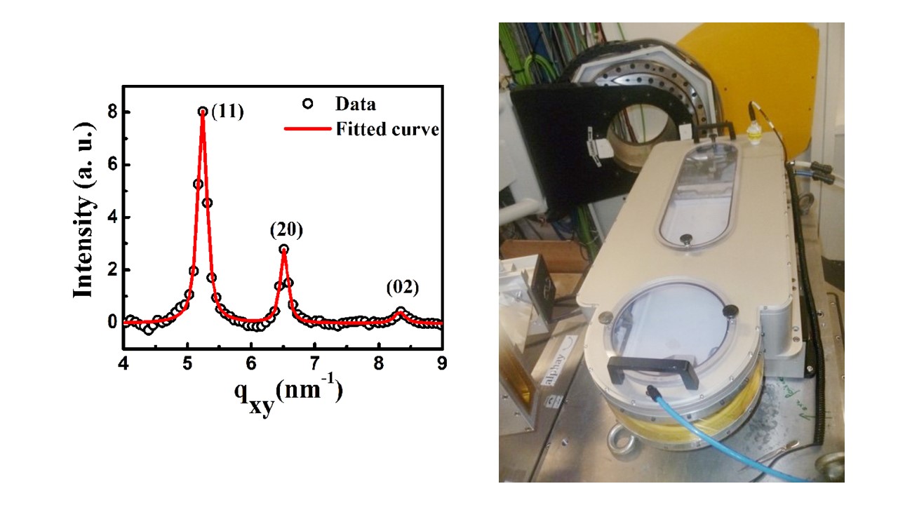
Left: Diffractogram measured on the 2D Cyclo-Ir crystals in-situ on the water surface showing the first 3 Bragg peaks (11), (20), and (02).
Right: Helium enclosure surrounding the Langmuir–Blodgett trough (white) adapted to X-rays, containing the layer under study at the water surface.d’étude.