Pearls: under the surface
Pearls are unique in jewellery: they are the only gem produced by living organisms. The evaluation of their quality, and consequently their price, is based on shape, colour, lustre..., i.e. their external characteristics. But what is under their surface? The study of their development carried out under the auspices of the Directorate of the French Polynesian Marine Resources research unit by scientists from the Museum national d’histoire naturelle, Sorbonne Université, ESRF and the SOLEIL Synchrotron NANOSCOPIUM beamline, reveals an unexpected diversity and complexity of structures and compositions. All these inner irregularities are the cause of surface defects, and permit the analysis of a still enigmatic phenomenon: the production of minerals by living organisms.
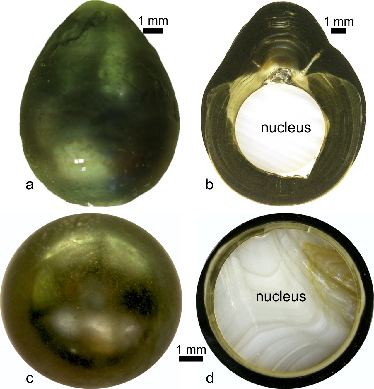
Invented in Japan in the early 20th century, cultured pearls are obtained by "grafting". A small fragment (the graft) of the organ producing the nacreous shell of a pearl "oyster" (Pinctada, not really an oyster) is inserted in a host of the same species. It is placed against a tiny sphere (nacreous nucleus, Fig. 1) previously inserted into the gonad of the "host" oyster. If the graft is successful (the loss rate is high), the graft survives, envelopes the nucleus and after approximately two years, the pearl is harvested and graded according to its qualities. A rigorous selection process is necessary, because, contrary to the usual descriptions, the nucleus has not simply been covered with a nacreous layer. Many pearls are irregular, but all reveal a complex process (Fig. 1). Detailed analyses of the internal structure of pearls and their composition gives direct access to the biomineralization process that takes place between grafting and harvesting.
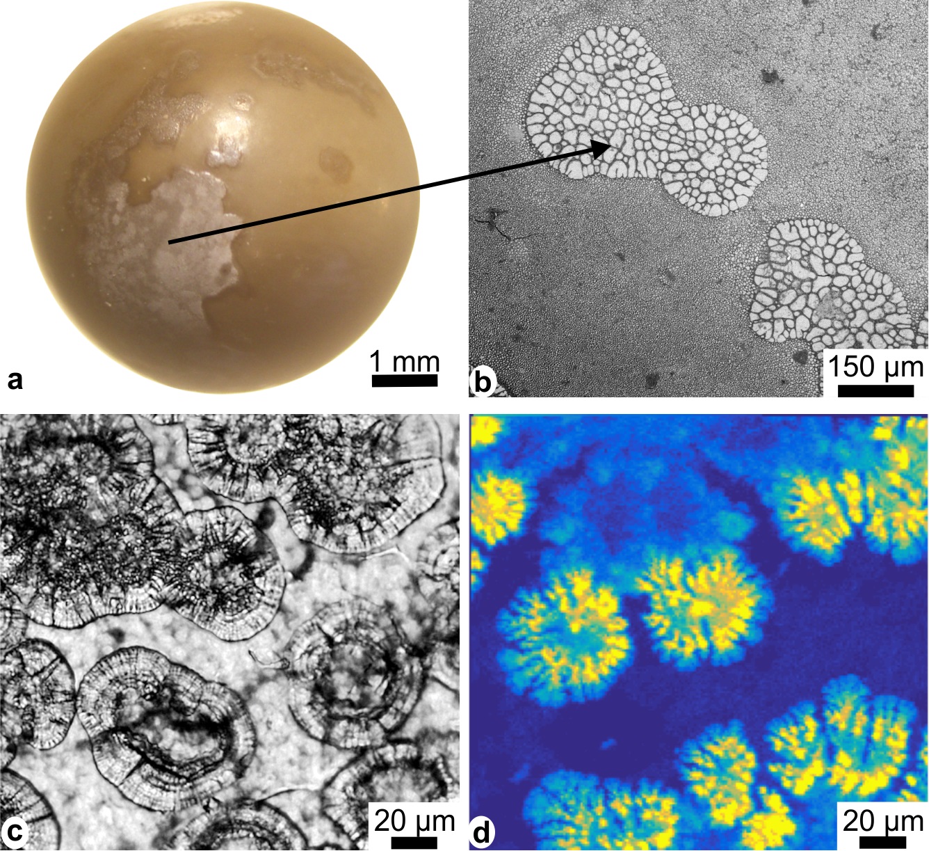
To observe sections of several hundred pearls has enabled the correlation of structural (micro- and nano-scale by microscopy and micro-tomodensitometry), mineralogical (Raman and infrared spectroscopy) and chemical (electron microprobe, micro X-ray absorption near-edge spectroscopy, nano X-ray fluorescence) data. It reveals that each pearl, whether round or not, has its own structure and composition. Abnormal structures (disrupting sphericity) are produced in the very first stages of the development of the pearl, indicating that the graft has significantly disrupted the production of the organic compounds controlling the bio-crystallization process. The disruption is sometimes so intense that in addition to the known structures in the shells (aragonitic nacre and calcitic prisms), unexpected new structures can be observed (aragonitic prisms for example are unknown in the shells). The irregularities are visible a couple of days after grafting (Fig. 2a, b). More or less large areas of varying shapes already contain mineral elements, while others are still entirely organic. Thanks to the nanometric resolution of NANOSCOPIUM, the presence of mineralized zones on samples that are one month old or less is clearly visible on the maps (Fig. 2c, d). The distribution of elements associated with calcium carbonate (calcite and aragonite) corresponds to both the radial and concentric arrangement of Ca.
So, even a round pearl is not a nucleus covered by a nacreous layer, as shown in the usual descriptions. It is now known that the quality of the graft (good health condition of the donor oyster, care in cutting the graft...) plays a major role, but a precise and direct correlation between these parameters, the inner structure and composition of the pearl, as well the quality of the surface is not yet unravelled. Indeed, cut pearls are not marketable. Finally, environmental parameters during the development of the pearl must also be taken into account.