How the Legionella bacterium hijacks a regulator of membrane traffic in infected cells
Legionella pneumophila, which causes the Legionnaire’s disease, escapes destruction by the immune system by hiding inside the infected cell. This camouflage strategy requires that the bacterium injects an arsenal of enzymes that take command of cellular pathways. One of these enzymes, AnkX, grafts a chemical compound to a major regulator of cellular traffic, thereby diverting it from its normal function. Snapshots of AnkX captured by X-ray crystallography revealed key aspects of how this reaction works.
The Legionnaire’s disease was named after a mysterious epidemic outburst that occurred in 1976 during an American legion meeting in Philadelphia. This severe pneumonia was shown soon after to be caused by Legionella, a bacterial pathogen that grows in ill-maintained air-conditioning and plumbing systems. It is transmitted through aerosol droplets to lung macrophages (a type of white blood cell), which are cells that normally kill pathogens. In France, 1200-1500 persons are infected each year, with a death rate of 10-15%.
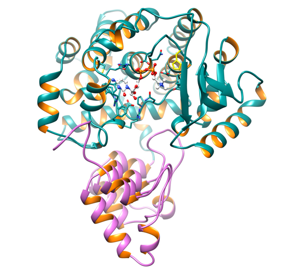
Legionella evades the immune system of the host by hiding in a membrane-bound structure, the legionnella-containing vacuole (LCV), where it multiplies in high numbers. To build, maintain and feed this vacuole in the infected cell, the bacterium injects an arsenal of enzymes that divert key host proteins from their normal functions. The Cherfils laboratory at the Laboratoire d’Enzymologie et Biochimie Structurales (LEBS, CNRS, Gif-sur-Yvette) solved the atomic structure of one of these enzymes, called AnkX. AnkX adds a small chemical compound, phosphocholine, to the small GTPase Rab1, a cellular protein whose normal function is to steer membrane traffic. This illegitimate chemical modification is pivotal for Legionella to reroute membrane traffic towards the LCV.
By using X-ray diffraction data collected at the PROXIMA 1 beamline, researchers captured snapshots of the chemical reaction catalyzed by AnkX. The crystallographic structures, combined with biochemical studies done in collaboration with the Roy laboratory (Yale University, New-Haven, USA), unveiled an unexpected enzymatic mechanism, and suggest that similar reactions occur with variations in other bacterial pathogens. These findings further our understanding of the molecular mechanisms whereby bacteria persist in the infected host.