Gum arabic reveals its secrets
Scientists of UMR IATE (Montpellier) and INRA (Nantes) have been studying gum arabic for the past few years, a bio-sourced material widely exploited in many sectors, such as the agri-food industry and particularly the wine industry, as well as the cosmetics and pharmaceuticals sectors and the paper industry. Experiments carried out on DISCO and SWING beamlines allowed them to determine a low-resolution 3D structure of the gum components, and helped them to better understand their function in the applications of gum arabic.
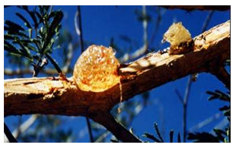
Gum arabic, or acacia gum, is a dry exudate obtained from the branches and stems of Acacia senegal (L) Willdenow, or Acacia seyal (Figure 1). In comparison, the European (E414) specification is slightly broader, since it encompasses all the species closely related to the Acacia (the pulse family).
The Gum arabic of interest here is very widely exploited in many sectors, such as the agri-food industry and particularly the wine industry, as well as the cosmetics and pharmaceuticals sectors and the paper industry, thanks to this material’s adhesive and emulsifying properties. In the specific case of winemaking, one of the main purposes of adding gum arabic before bottling is to stabilise the colouring substances in red wines and reduce the risks of ferric or copper casse in white and rosé wines. Gum arabic therefore constitutes an excellent colloidal stabiliser, with no allergenic properties. Very few studies, however, have examined the structure of the constituents of gum arabic at the same time, and even fewer have looked at the interactions involved in the colloidal stabilisation of wines.
The IATE UMR (mixed research unit) at Montpellier and INRA Nantes have been working for the past few years on the fractionation of gum arabic, on determining the structure of the majority components obtained by this fractionation, and the interactions involved when gum arabic is in the presence of proteins.
Fractionation of gum arabic
Gum arabic consists of polysaccharides rich in galactose and arabinose, and a small proteic fraction associated with these polysaccharides. It is highly soluble in water, up to concentrations in the region of 500 g/l. Even so, to understand the relationship between structure and function of gum Arabic, and its functional properties in particular, the complexity of gum Arabic can be dealt with by separating the various polysaccharides of which it is made. Thus, in the first instance, based on the knowledge acquired, we have fractionated the gum arabic via a hydrophobic interaction chromatography phase, in order to separate the species that are present according to their different degrees of hydrophobicity. In this way, we were able to show that gum arabic is a continuum of molecular species that differ from each other by their mass, charge, and sugar/protein ratio (1). The three majority species arising from fractionation are arabinogalactan (AG) (88%), arabinogalactan-protein (AGP) (10%) and glycoprotein (GP) (2%). Note that these three species may be represented in variable proportions according to the sampling date or the age of the trees, for example, and that the AGP species can have significant structural variations in terms of molecular mass. This AGP species in particular is strongly involved in the stabilisation of consumable emulsions such as acidified drinks (e.g. sodas). It is not always easy, however, to eliminate the natural variability of gum arabic without homogenising the batches of gum arabic based on different harvests or using a treatment process under a controlled atmosphere and temperature developed and patented by a Japanese company (2) to make the AGP structure uniform and give the gum arabic uniform emulsifying properties.
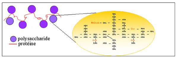
The majority fractions purified in this way allowed us to perform structural studies to show the conformation in space of the three macromolecules that make up gum arabic: arabinogalactan (AG), arabinogalactan-protein (AGP), and glycoprotein (GP). The generally accepted structure of arabinogalactan protein (AGP) to date is the one presented in Figure 2, called the “wattle blossom model”, where the polysaccharide blocks (purple circle) are covalently bonded to the protein chain. Moreover, each polysaccharide block essentially consists of arabinose and galactose patterns bonded to each other to form a complex and multi-branch structure. The polysaccharide blocks also contain charged patterns (uronic acids), which can be involved in colloidal stabilisation.
Determining the structure of the gum arabic fractions
Because of their large size, these macromolecules cannot be crystallised to determine their structure at a high resolution. That is why we have used methods other than x-ray crystallography to determine a low-resolution three-dimensional structure. From our knowledge of the three-dimensional structures, we therefore hope to be able to identify the relationship between structure and function for each of these macromolecules, and ultimately adapt the formulations for the different applications of gum arabic, particularly in terms of stabilisation.
The structure of arabinogalactan (AG) could therefore be established via the scattering of the radiation and a calculation to reconstruct the most probable three-dimensional structure. In parallel, microscope observations have been used to compare images to the computed model (Figure 3) (3). The characteristic dimensions of the structure of AG, comparable to a thin disc, are 20 nm long and 2 nm thick.
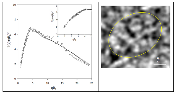
The three-dimensional structure of arabinogalactan-protein (AGP) could not be established because of the presence of two species in this purified fraction. Nevertheless, the comparison of two structural methods, radiation scattering and microscopy, have allowed us to reveal a conformation comparable to a thin triaxial ellipsoid with maximum dimension equal to 64 nm, whose internal structure would be similar to the wattle blossom model described in the literature (Figure 4) (4, 5).
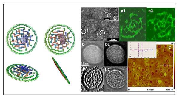
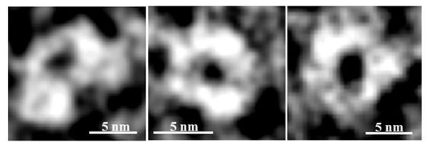
Likewise, the structure of glycoprotein (GP), the most recent work carried out at SOLEIL at SWING and DISCO beamlines has revealed oligomers (an assembly of monomers) that are thin, with an ellipsoidal conformation. In addition, these oligomers appear to be the result of an assembly of sub-units in the form of rings (Figure 5). These sub-units are characteristic of hydroxyproline-rich glycoproteins (amino acid onto which polysaccharide blocks are grafted).
Can this knowledge of the structures of the three fractions help to better understand their function in the applications of gum arabic, of which there are now so many?
Relationships between structure and function and consequences on the functional properties of gum arabic
Firstly, the abnormally low viscosity of gum arabic should be linked to the small size (D = 20 nm) and the very low density of the main fraction (AG) (d = 1.007) and the AGP fraction (d = 1.005). The dimensions of the gum arabic fractions testify to the very thin configuration (e < 2 nm) of macromolecules with a high molecular mass. These very low thicknesses are certainly the cause of the very strong adhesive capabilities of gum arabic, both on solid surfaces (e.g. paper) and liquids (e.g. in the case of emulsified drinks). On the other hand, the adhesive capabilities of gum would not appear to be related to the majority AG fraction because of its strongly hydrophilic polysaccharide chemical nature. The adhesive properties of gum arabic seem to be contained in the other two minor fractions, AGP and GP. In particular, the protein part contained in AGP and GP, which is hydrophobic and amphiphilic, could make these fractions more efficient in terms of adsorption at solid and liquid interfaces. Because polysaccharide part forms a highly hydrophilic external layer, its function would be much more involved in colloidal stabilisation by steric repulsion.
References:
(1) D. Renard, L. Lavenant, M-C. Ralet, C. Sanchez (2006) Biomacromolecules 7, 2637-2649.
(2) H. Hayashi (2002) Enhancement method of gum arabic under the atmosphere of 30-100% of relative humidity at over 40 °C. Patent Japan 2002 130212; PCT-JP02/08144; WO 03/093324A1; US 2005/0158440.
(3) C. Sanchez, A. Lapp, C. Schmitt, C. Gaillard, E. Kolodziejczyk, D. Renard (2008). Biophysical Journal 94, 629-639.
(4) D. Renard, C. Garnier, A. Lapp, C. Schmitt, C. Sanchez (2012). Carbohydrate Polymers 90, 322-332.
(5) D. Renard, C. Garnier, A. Lapp, C. Schmitt, C. Sanchez (2013). Carbohydrate Polymers 97, 864-867.