Discovery of the oldest macroscopic planktonic eukaryotes - Contribution of the NANOSCOPIUM beamline
The origin of eukaryotic* cells is a major challenge in evolutionary biology and has been sharply debated within the scientific community for decades. An international team coordinated by Abderrazak El Albani (IC2MP - Université de Poitiers/CNRS) has discovered the oldest fossils of eukaryotic protists, which lived in seawater (plankton) 2.1 billion years ago. These fossils were discovered in the famous Moulendé deposit in Gabon, which had already provided the oldest multicellular "Gabonionta" organisms, shedding new light on the early emergence of eukaryotes.
This discovery pushes back the time-frame for the emergence of eukaryotic organisms by over 300 million years.
A few years ago, Abderrazak El Albani's team at the “Institut de chimie des milieux et matériaux“ in Poitiers, France, discovered the oldest fossils of multi-cellular organisms in Gabon, known as "Gabonionta". Thanks to this deposit, located in the Franceville basin, the date of appearance of multicellular life on Earth was pushed back by around 1.5 billion years, from 600 million to 2.1 billion years ago. Researchers had also shown that this formidable biodiversity, concomitant with a peak in atmospheric oxygen concentration, had developed in a calm, shallow marine environment. It was within the same geological formation that the IC2MP team discovered the existence of fossils of macroscopic protists up to 4.5 centimetres in size.
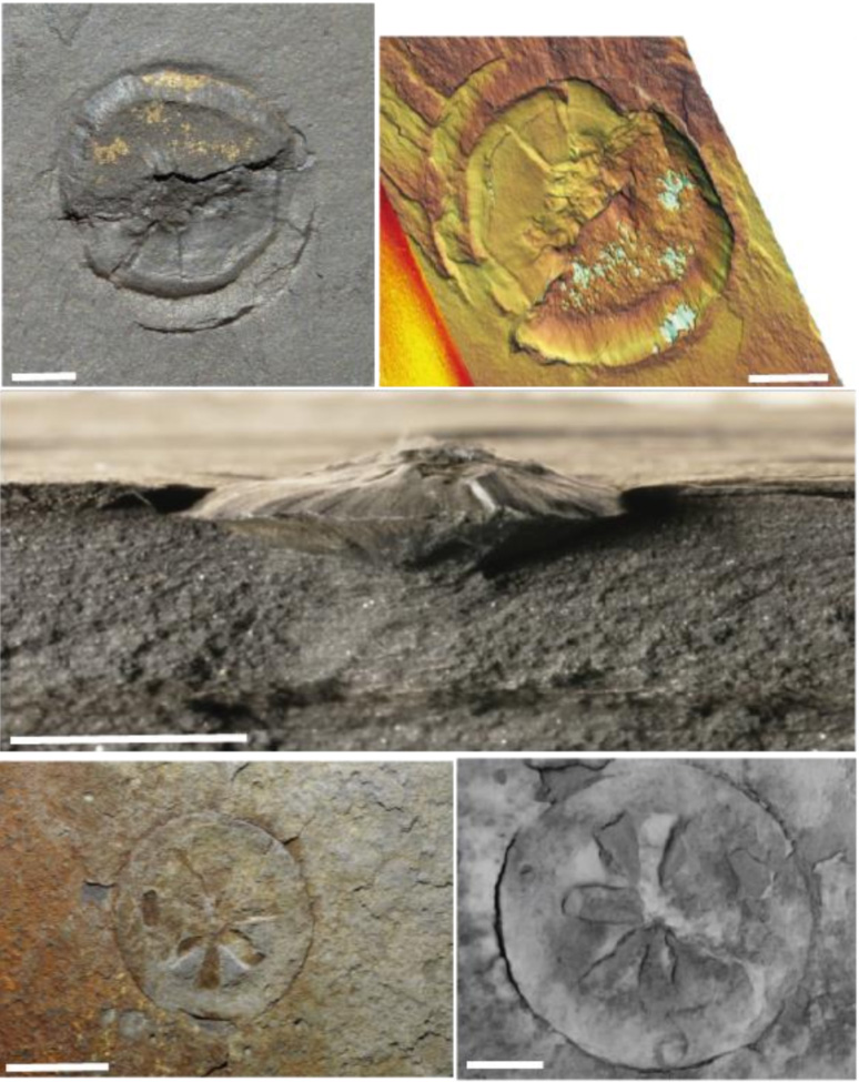
They seem to have been living in the water, not on the seabed. In this primitive marine ecosystem, some eukaryotic organisms were already biologically sophisticated enough to live planktonically. This result was achieved thanks to the use of zinc as biomarker, considered as an indispensable element for biological metabolism. The data revealed that these fossils contain around twice as much zinc as the sediment in which they are found. What's more, the zinc isotopes within these fossils show an enrichment in the lighter isotope compared to the same sediment. The behaviour of zinc is of great interest as it is a bio-essential micronutrient, a component of several metalloenzymes performing key biological functions in eukaryotic cells.
Thus, the cellular zinc demand is highly dependent on the size, organization, complexity, functionality, and metabolism of the cell. In this respect, an increase in the size of these organisms could be correlated with an increase in zinc availability. These data highlighted the fundamental role of zinc as a biogeochemical marker of biogenicity.
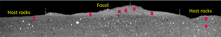
In parallel, the study by scanning X-ray fluorescence nano-imaging on SOLEIL's NANOSCOPIUM beamline revealed the nanoscale spatial distribution of zinc within these specimens, as well as its co-localization with organic matter. Moreover, X-ray full field micro-tomography, a non-destructive imaging technique, shed light on their 3D morphostructure. The obtained results showed unambiguously that these lenticular structures have an internal compartmentalization. Thus, these imaging techniques provided further confirmation of the biological origin of these fossils.
What did these living creatures actually look like?
They may have been similar to large protists capable of floating in the water column. When these organisms aggregate fine clay particles, their density causes them to fall randomly onto the sediments forming the seabed. Until now, the oldest recognized planktonic eukaryotic protists were dated at 570 million years, which seemed to be supported by estimates using the molecular clock**. This new discovery highlights a biological innovation that raises new questions about evolutionary history: did advanced planktonic life forms already exist 2.1 billion years ago?

Contribution of the NANOSCOPIUM beamline
Scanning XRF imaging has been performed by fast continuous scanning (FLYSCAN) at NANOSCOPIUM on mm2–sized regions within representative samples. This provided elemental distribution maps of S, K, Ca, Ba, Mn, Fe, Ni, Cu, Zn, Ga, Ge, As, Se, Pb, Sr, and Zr within lenticular-shaped fossils and the embedding sediment (host rocks), as illustrated in Figure 3a & b.
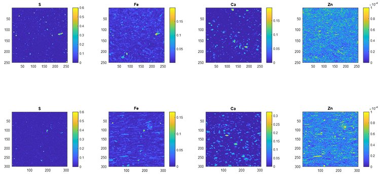
The large number of pixels, 20 000-90 000 per image, measured in each scan allowed thorough statistical comparison between the chemical composition of the fossil and host rock. For example, as the distribution maps of S, K, Ca, and Mn do not show any significant co-localization with Zn, it can be concluded that Zn is not linked to S, K, Ca, or Mn-containing mineral phases whether in the fossil or host rocks. Moreover, high spatial resolution XRF imaging was crucial to identify high Zn-containing outliers, which are present in only ~3.5 % of the total number of pixels as micro-meter sized mineral phases. The rare presence of these Zn-bearing micro-minerals results in a negligible, < 10 %, increase of the measured mean Zn concentration of any chosen sample area. This finding is crucial for the reliable comparison of the Zn content of the fossil filling and host rocks by bulk analytical techniques.
* Organisms composed of one or more cells, characterized by the presence of a nucleus and organelles delimited by cell membranes.
** The principle: exploit the variations observed between two species in similar regions of their DNA to estimate the time elapsed since their last common ancestor lived.