Breast cancer: link between inflammation and microcalcifications
In France, each year, breast cancer represents one third of new cancer cases in women. The incidence of this cancer is increasing, but fortunately with a decreasing mortality rate. Despite this situation, diagnostic tools have not evolved much and the first tool of detection remains the mammography.
The presence of microcalcifications in breast tissue and their observable distribution on mammograms are used as indicators of the presence/absence of cancer. However, knowledge about the relationship between the presence of microcalcifications and cancer severity is patchy. Few systematic studies have been undertaken to understand the interactions between calcification formation, tumor presence, inflammation and extracellular matrix remodeling.
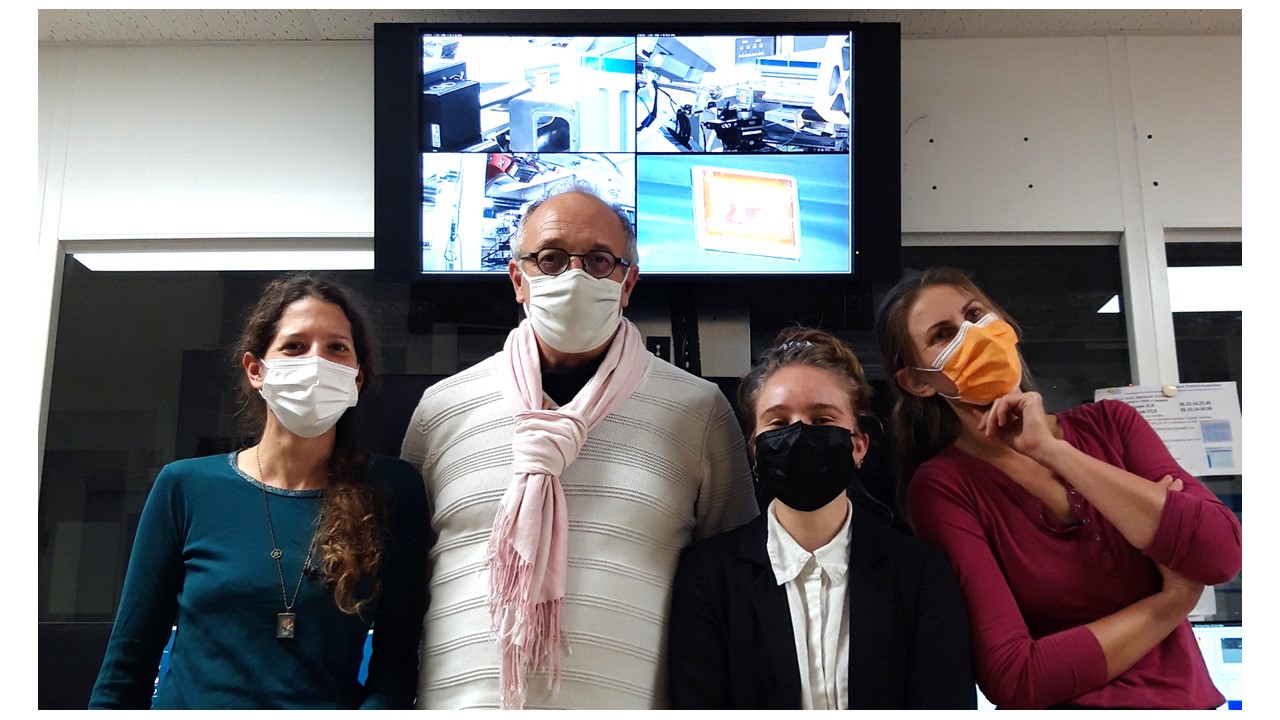
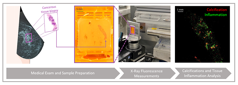
In this context, researchers from the Institut de Chimie-Physique (ICP), present in October on the DiffAbs beamline (see picture below), wish to evaluate the link between the presence of microcalcifications and tissue inflammation in cancer patients.
Zinc is considered a biomarker of tissues with inflammation, so they proposed to localize calcium1 and zinc cations in tissues by X-ray fluorescence microscopy. To conduct this study, breast tissue biopsies from patients with non-invasive and invasive2 carcinoma will be explored.
Initial tests performed in October on a tissue biopsy from a patient with non-invasive carcinoma (see figure 1) have demonstrated the feasibility of the experiments and have already revealed very promising results: there appears to be partial co-localization of the calcium and zinc fluorescence signal, which differs from observations made for other pathologies involving calcifications.
To confirm this result, the ICP scientists have applied for beamtime to perform a statistical study on a large number of biopsies. They hope to clarify the mechanisms of pathogenesis of micro-calcifications as well as their role in cancer severity.
The project is realized in collaboration with Dr. Maguy Cherfan from René Dubos Hospital in Pontoise.
1 - Calcium: Element present in the micro-calcifications
2 - Cancer localized in the breast ducts (non-invasive carcinoma), with spread of cancer cells into the surrounding tissues/organ (invasive carcinoma)