Breaking up molecules with UV radiation: a new activation method for tandem mass spectrometry
Tandem mass spectrometry is of central importance in analytical and structural chemistry. This technology involves breaking up the molecules being studied to deduce information about their initial structure by analysing the way in which they fragment.
The quality of the information obtained depends on the method employed to achieve this fragmentation. One of the tools used to break the chemical bonds between molecules is light. In collaboration with INRA, one of SOLEIL’s scientists has identified a part of the electromagnetic spectrum that is particularly effective in breaking up molecules and developed a new source for the fragmentation of molecules for mass spectrometry.
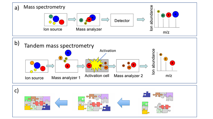
Since the advent of the ‘modern’ methods of ionisation (such as electrospray and MALDI), mass spectrometry has become one of the main technologies used in the analytical sciences. A mass spectrometer comprises (figure 1 a):
- an ionisation source capable of changing molecules into the gaseous phase and ionising them,
- a mass analyser which separates the ions based on their mass-to-charge ratio (m/z),
- a detector which records the passage of ions, dependent on their m/z.
A mass spectrum is a representation of the abundance of ions as a function of their m/z ratio. Based on measurement of these m/z ratios, it is in particular possible to calculate the molar mass of the species studied.
A variant of the ‘single’ mass spectrometer exists which yields more information than this single variant. Indeed, the addition of an additional mass filtering stage, as portrayed in figure 1 b), is used to isolate one of the ions obtained during the first stage, to cause it to absorb energy (referred to as the activation stage) and then to observe the fragment ions resulting from this energy absorption.
This method is referred to as tandem mass spectrometry MS/MS or also MS2. When several selection and activation stages are performed in succession, the process is referred to as MSn.
The interest in MS/MS is the ability to be able to reassemble the precursor ion structure by analysis of the fragmentation profiles, as illustrated in figure 1 c).
Currently, this technique is primarily used for the sequencing of biological macromolecules (proteins, etc.).
How can the activation stage be improved?
In tandem mass spectrometry, the ion activation stage is crucial because it is largely responsible for the nature of the fragmentation products. The most widely used method is collision induced dissociation (CID). It involves inducing inelastic collisions between the ions studied and an inert gas (for example N2 or Ar) in a collision cell.
CID, the standard method offered in all commercial MS/MS devices, entails slow heating of the ions. The multiple collisions undergone by the ions, gradually increases their internal energy until a dissociation threshold is reached. The result is that the weakest bonds are broken. Nevertheless, this is not always desirable, especially when the species of interest have labile chemical groups such as post-translational protein modifications (phosphorylations or glycosylations) or in the case of fragile assemblies such as non-covalent complexes formed between biological molecules and their ligands. The slightest activation by CID causes these complexes to dissociate with the result that the information about the molecule/ligand interaction sites, which is of primary importance in multiple areas (mode of action of drugs, molecular recognition, etc.), is lost.
In spite of the ease with which CID is implemented and its relatively low cost, these limitations have led to the development of alternative methods.
The solution: light
One way of rapidly supplying a great deal of energy to an ion is to cause it to absorb photons. The more energetic the photons, the greater the quantity of supplied energy. This idea was implemented by coupling powerful lasers, which emit radiation in the ultraviolet (UV) and vacuum ultraviolet (VUV) ranges, to the ion traps. Activation of ions by UV or VUV has specific characteristics which are complementary with other methods of activation such as CID.
Nevertheless, the emission wavelengths are limited: commercial lasers can only access the lowest excitation states of the electrons. The obvious question then arises of whether it is possible to use more energetic radiation.
To enable investigation of the higher energy ranges, an ion trap mass spectrometer was successively coupled to the DISCO and DESIRS beamlines. Indeed, synchrotron radiation is a bright and tunable source (possibility of choosing a given wavelength or energy) over a very wide range of energies. This enables investigation of fragmentations induced by light absorption across the entire electromagnetic spectrum.
It was possible to study the main families of biological molecules: sugars, proteins, etc. and evidence of new fragmentation paths was provided.
Hence it was found that the most effective range of energy for tandem mass spectrometry is centred on 20 eV (~ 60 nm wavelength) in a range referred to as the extreme ultraviolet (XUV). Indeed, this range corresponds to the absorption maximum of the majority of chemical species and it creates a particular fragmentation mode referred to as dissociative photoionisation (DPI).
Now, it turns out that this range of wavelengths, although outside the range of commercial lasers, is within the range of discharge lamps.
From the synchrotron to the laboratory
A scientist working on the DISCO beamline, also affiliated with INRA, exploited the results obtained using the light from the SOLEIL synchrotron to install a discharge lamp on a commercial mass spectrometer. The system was successfully tested on model molecules (sugars, peptides, etc.). The fragmentations induced by the radiation emitted from the lamp – a helium discharge lamp was found to be the most effective – reproduces the findings obtained using the synchrotron radiation.
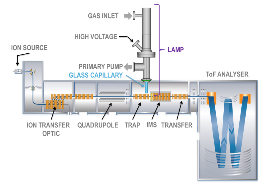
A new activation method for tandem mass spectrometry, based on XUV radiation produced by a rare gas discharge, has thus been developed.
Therefore, this technology makes available radiation which is capable of photoionisation of ions produced by electrospray or by MALDI to induce their fragmentation. The fragmentation profiles obtained are complementary to other methods and, in certain cases, the obtained information content is of better quality.
Moreover, this inexpensive method, which is easy to implement with commercial equipment, is not subject to the safety requirements imposed on laser systems. Therefore it will be an effective competitor amongst the arsenal of activation methods available to researchers.