HERMES
HERMES beamline is dedicated to X-Ray microscopy. It is made up of two branches, each one having a microscope available: STXM (Scanning Transmission X-ray Microscopy) and X-PEEM (X-ray PhotoEmitted Electron Microscopy). Both microscope are complementary and will be running on alternatively.
HERMES is a phase III beamline dedicated to X-ray microscopy. It combines two types of microscopy. The first one is a Photon-Photon microscopy: Scanning Transmission X-Ray Microscopy (STXM). The second type is Photo-Electron microscopy: X-Ray Photoemitted ElectronMicroscopy (X-PEEM). This original approach offers to users two very complementary techniques, each one being adapted to specific sample environments.
The STXM essentially enables to probe the sample volume’s properties, with depths of analysis around few hundred nanometers. Besides, X-PEEM is a surface sensitive technique adapted to an ultra-vacuum environment. It mainly enables to scan the surface’s first few nanometers.
Both techniques carry out X-Ray spectroscopy as a contrast method, in this case a chemical contrast.
Alongside, other contrast means can be added by making use of the spectroscopic techniques’ specificities. Thus, using circular and linear polarization of light gives an access to magnetic domains, ferromagnetic and antiferromagnetic (XMCD and XMLD)…
Finally, both methods can be used not only to get images of samples, but also to realize some measurement in local spectroscopy (XAS, XANES, XPS, ARPES…) at a nanoscopic scale.
Beamline news
All HERMES news
SOLEIL Highlights 2025
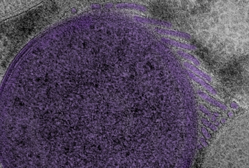
Contribution of the HERMES beamline to the study of "Tubenets”: Network-like structures (...)
Team


Technical data
- Description
-
HERMES beamline is installed on a medium section (IM9). The beamline covers the energy range going from 2.5 eV to 2,5 keV, to meet the requirements of the scientific programs. The beamline starts right below the Si2p-edge, covers the K-edge of light elements (C, N, O...) , L-edge of transition metals, M-edge of rare earth and eventually the K-edge of Si, S and P. Furthermore, the beamline enables to work with the variable linear polarization (horizontal to vertical), in addition to circular and elliptical polarization.
To enable this, the beamline has two undulators, Apple-II type : HU-64 (1.7m, 25 periods) for the low energy, covering the 70-600 eV range. HU-42 (1.8m, 42 periods) for the high energy range, going from 0,5 to 2,5 keV.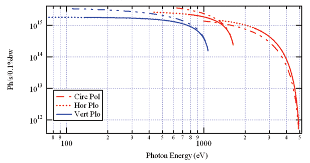
Figure 1 : Flux calculé pour les onduleurs HU64 (bleu) et HU42 (rouge)
Figure 1: Calculated flux for aperiodic HU64 (blue) and HU42 (red) undulatorThe beamline is demarcated into five sections:
- Mirror chamber M1: It deflects the beam and absorbs most of the thermal load.
- Monochromator (VLS-VGD and multilayer): It enables to combine the two types of monochromators so that the whole energy range is covered with a good efficiency and a high resolution, keeping the same optical path.
- Mirror deflection chamber M4,M5: The M4 and M5 mirrors deflect the beam respectively to X-PEEM and STXM branch.
- X-PEEM branch: The beam is focused using Kirk Patrick-Baez (KB) mirrors to obtqain a beam of size 7x25µm² on the X-PEEM sample position.
- STXM branch: This branch contains a slit system determining the secondary source for the ZP lenses and the STXM microscope.
Several beam diagnostic systems are installed throughout the line.
- Energy range
-
70 – 2500 eV
- Source
-
Undulator HU64 : Low energy
Undulator HU42 : High energy
- Flux at the source
-
< 1015 phs/s/0.1% b.p. on the whole range
- Polarisation
-
Variable polarisation : circular and linear
- Resolving power
-
E/∆E > 5000 on the whole energy range
- Optics
-
• Double mirror in chicane mode (2 angles of light for the harmonic rejection)
• Double monochromator:- PGM in mode VLS-VGD (600 et 450 lines/mm) or variable deviation mode (Petersen) for low energy
- Optical multilayer grating for high energy
• X-PEEM branch : Refocusing mirrors KB bender
• STXM branches : ZP lenses
- Beam size
-
• X-PEEM branch : 7µm x 25µm (VxH)
• STXM branch : Φ300µm on the ZP, 25nm on the sample
- Flux on sample
-
• X-PEEM branch: > 5.1012 Ph/s
• STXM branch: > 1.1011 Ph/s
- Sample environment
-
• X-PEEM branch:
- Ultravacuum or controlled environment (P<1.10-5mbar)
- In-situ preparation possibility: Evaporation, Ar etching, heating, cleavage...
- Complementary in-situ analysis: LEED, LEEM, Auger AES, Kerr.
- Sample temperature : 150-2000K
• STXM branch :
- Secondary vacuum, controlled environment (<1barr)...
- Magnetic field < 200 Oe
- Sample temperature : 150-600K
- Wet Cell, Microfluidic...
Scientific Opportunities
| Scientific theme | Methods |
|---|---|
| Magnetism | This program is primarily aimed at nanostructures and magnetic material study. These requested studies pair up imaging experiment as well as local spectroscopy. This thematic benefits from circular and linear polarization from synchrotron radiation. The dynamic range of studies (sous champ, induced current…) also fit the expectations of the users’ community. There is also a big interest for temperature measurements, especially to adapt to Curie’s and Neel’s temperatures (150K-1500K). Several subjects have been developed on the beamline: spin electronic, magnetic tunnel junction, exchange coupling anisotropy, nanostructuration and size effects, multiple ordered system (multiferroics…), antiferromagnetic oxides, magnetization dynamics… • XMCD-XMLD imaging and spectroscopy |
| Nanostructure | There are also big needs in this domain, especially concerning local spectroscopy developments, XPS, XAS, XANES… Overall, this thematic covers the study oflow dimensionality effects and auto-organization on properties and electronic structure of nanomaterials. • Traces element analysis |
| Soft Matter | This program will essentially benefit from the unique possibilities of measurements in water window (K-edge of carbon, nitrogen, oxygen…). This program also requires special sample environments to study hydrated samples ("wet cell", "microfluidic"…). The use of beam polarization also enables to detect molecular orientation on different surfaces. • « wet cell », micro-fluidic |
| Surface/Interface | This thematic is exclusively developed on the X-PEEM microscope. The LEEM (Low Energy Electron Microscopy) and X-PEEM enables to study the structure, morphology, chemistry and electronic properties of surfaces and interfaces in-situ and on the same sample . Several issues can be treated: layers growth, surfaces catalysis and chemistry, magnetism, semi-conducting surfaces… • Local imaging and spectroscopy |
| Earth and Environment Science | This program combines STXM’s spectroscopic aspects of STXM with aspects of mineralogy, and will fully benefit from the energy range of the beamline. Several issues under this themeare common with other themes, such as bio-mineralization, toxicity, chemistry and nanostructure. A particular attention is paid to local spectroscopy aspects, especially at the carbon edge. • Local imaging and spectroscopy |
| Biology and medicine | This program is relatively wide and relies on structural characterization (cellular core, bacteria, micro-organisms...) identification and chemical co-localization and configuration, as well as on labelling and translocation aspects. This program should significantly benefit from cryogenics and micro-fluidics. • Cryogenics |
As a consequence to the microscopes complementarity, HERMES’ scientific program is wide and affects several communities. It is defined in the beamline’s APS and has been approved by SOLEIL’s scientific committee. It covers the following themes (for more details, refer to the beamline’s APS) :
Methods
Scanning Transmission X-Ray Microscopy
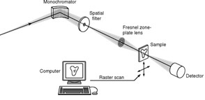
STXM microscopy is a scanning microscopy technique. It is based on an extreme focalization of the X-beam, using focalizing Bragg-Fresnel lenses (Zone Plate). Thus, the beam’s size determines the resolution. The beam is focalized and scans the studied zone of the sample. The transmitted beam is detected with different detectors (CCD camera, PM Scintillator…)
X-Ray PhotoEmitted Electron Microscopy
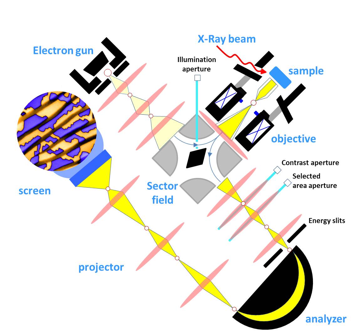
X-PEEM is an indirect microscopy technique. An X-PEEM microscope is based on the same principle as a low energy scanning electron microscope. In addition to the absorption of photons, emitted photoelectrons from the sample are collected, dispersed, analyzed and eventually projected on a detector, using an electron optical column. In this way, we get a magnified image of the working area on the detector, rebuilt from the photoelectrons.
Schematics
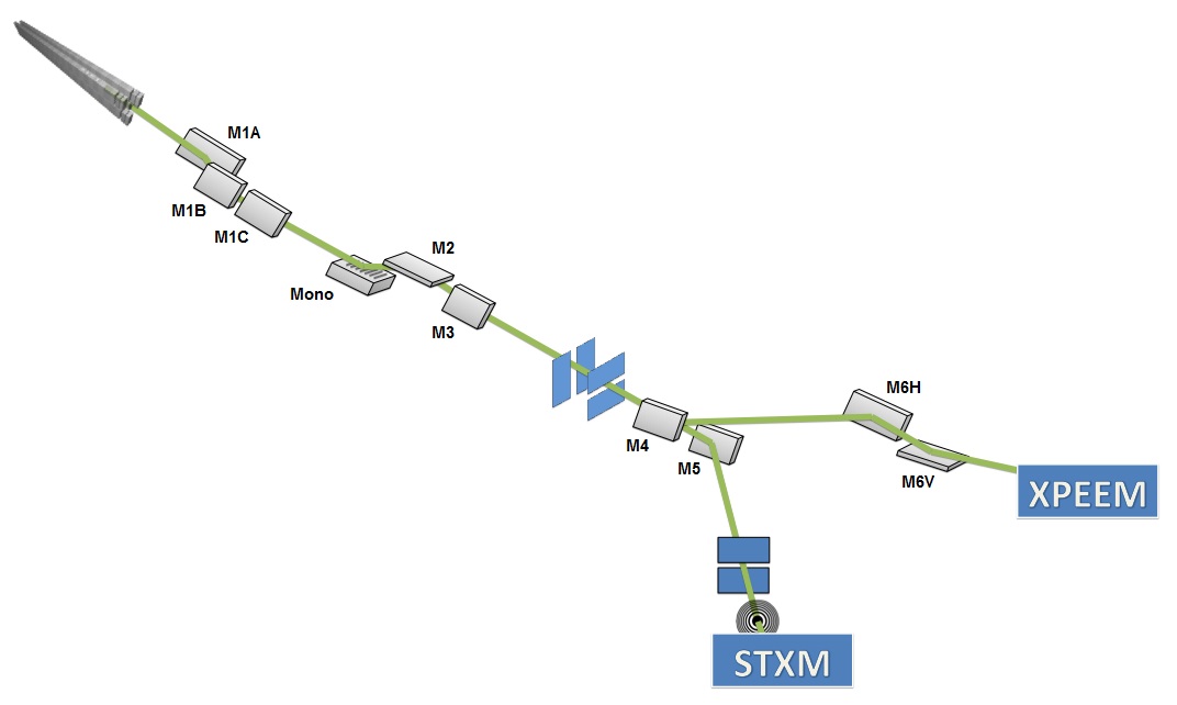
Figure 2 : HERMES's optics
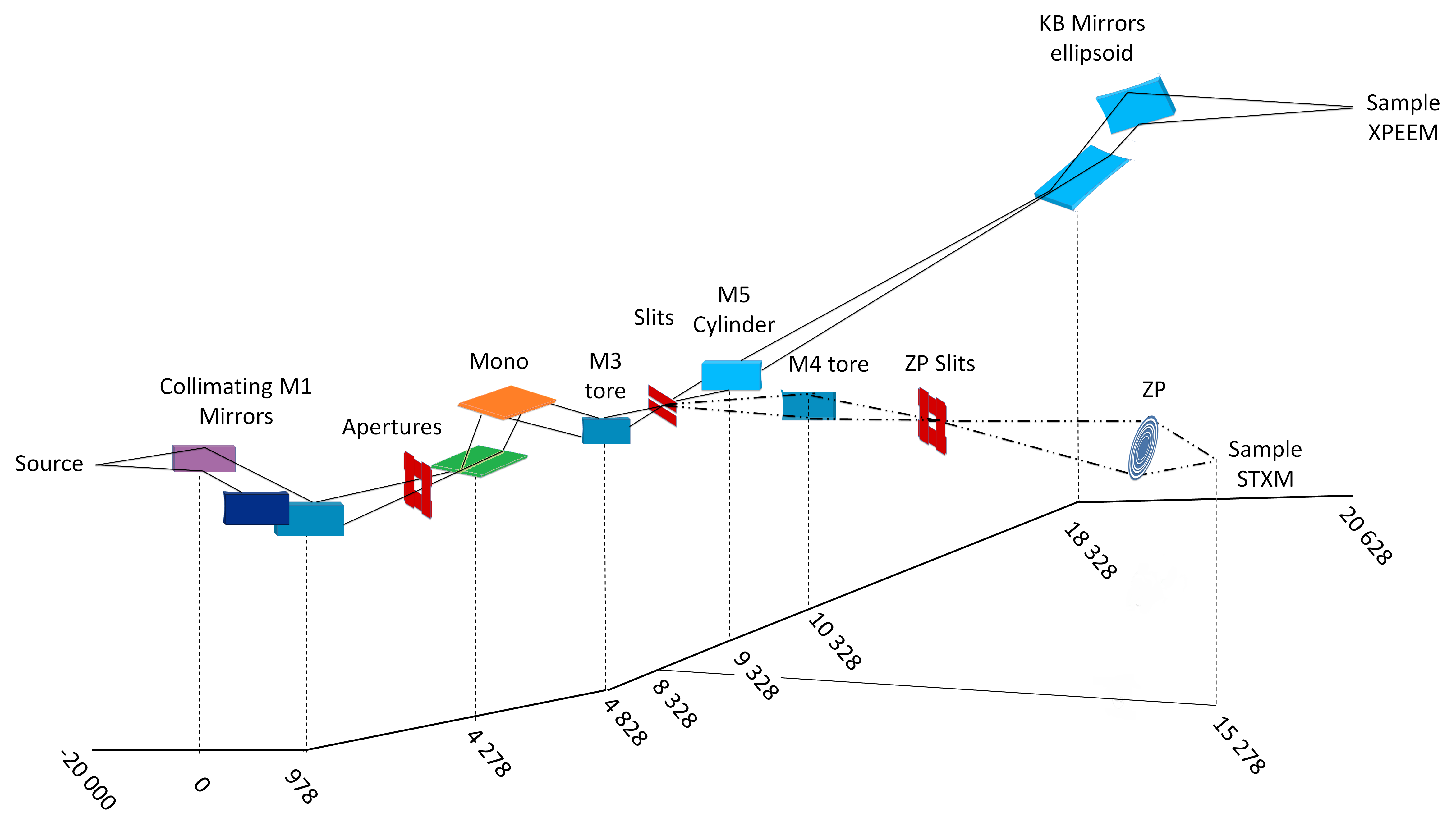
Figure 3 : HERMES' Implantation
The HERMES beamline welcomes users since november 2015 for the X-PEEM branch and october 2016 for the STXM branch. Both microscopes receive beam alternatively. Each microscope is dedicated to specific scientific domains with complementary sample environement and preparations.
Samples
X-PEEM samples:
The samples characterized by X-PEEM at HERMES are mounted on ELMITEC cartridges and are therefore submitted to the associated spatial constraints. Hence the thickness should not exceed 4 mm and the lateral dimansions 1x1 mm² or 1.2 cm diameter.
It is more desirable to have a conductive sample (or capping layer) to avoid charging effects.
.
Specifications
XPEEM preparation chamber:
- Heater stage (up to 2000 K)
- Ar+ ion gun for sputtering
- Various gas inlets (e.g. oxygen)
- Sample carrousel (up to 6 samples)
XPEEM microscope environment:
- Ultra-high vacuum environment (10-11 mbar)
- Motorized (X,Y) translation (± 2500 µm)
- Motorized azimuthal rotation (0 – 360°)
- Temperature control (115 K – 2000 K)
- In situ current injection / magnetic field foreseen
- In-situ electric polarization foreseen
- In-situ evaporation with e-beam & Knudsen evaporators
- Hg lamp (4.9 eV), He lamp foreseen (21.6 eV)
XPEEM microscope specifications:
- Full field imaging (fast acquisition time)
- Fields of view from 50 µm to 2.5 µm
- Resolution in LEEM mode < 10 nm
- Resolution in PEEM mode < 30 nm
- Analyzer resolution: 100 meV (50 meV ultimate)
Characterization techniques catalogue:
- Surface sensitive (< 10 nm probed depth)
- Morphology and crystal structure with LEEM and µ-LEED
- Local absorption spectroscopy using XAS and XANES
- Local electronic structure with XPS, RPES, ARPES and PED
- Imaging of ferro/antiferromagnetic domains using dichroism (e.g. XMCD, XMLD, XLD)
Useful information
HERMES X-PEEM Images Analysis using IgorPro:
- Download the X-PEEM Images Analysis IgorPro procedure file: download
- Copy this procedure file in the folder ...\WaveMetrics\Igor Pro Folder\Igor Procedures or...\Documents\WaveMetrics\Igor Pro 6 User Files\Igor Procedures
- The X-PEEM Images Analysis panel is now usable in IgorPro
You can use the short user manual of the X-PEEM Images Analysis procedures: download
manual_xpeem