Radiotherapy : a new method to localize unlabeled nanoparticles in tumor cells
To improve radiotherapy techniques, several studies have looked at the possibility of adding nanoparticles, especially when treating certain aggressive tumors, such as glioblastoma cells (the most common brain tumor). However, these particles are difficult to locate precisely inside the cells. Until now fluorescent markers were used to follow the path of these nanoparticles. The problem was that these markers could directly influence their location, thus skewing the study. A group from the Institut des Sciences Moléculaire d'Orsay (UMR 8214 CNRS-UPSud) has recently proposed a combination of techniques developed at the Institut Curie (Orsay) and the SOLEIL synchrotron DISCO beamline (Gif / Yvette) to track the position of unmarked nanoparticles (synthesized by researchers from the Institut Lumière Matière, Lyon 1), used to amplify the destruction of human tumor cells. The results are published in the journal Cancer Nanotechnology.
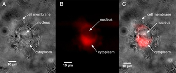
Radiotherapy remains a frequently used treatment for some very aggressive tumors: radiation (X-ray, gamma or ions) targets tumors to destroy them, with the risk of collateral damage to adjacent healthy cells. In order to optimize radiation therapy, the scientific community has long been interested in the effects of nanoparticles (NPs). These are used in diagnosis or in therapy, as their presence in cells increases the radiation effect. However, their entry, exact location and how NPs act in tumor cells is often unknown. Now, for the first time, a group has used synchrotron radiation deep UV (SR-DUV) microscopy available on theDISCO beamline of the SOLEIL synchrotron in order to improve understanding of these processes. The researchers observed human glioblastoma cells in the presence of Gadolinium-based nanoparticles (GdBN), known to be effective in diagnosis and therapy, but also for their low toxicity. Without the need to label the NP surface, the technique has helped locate nanoparticles in cells due to their autofluorescence. These are found to be localized in the cytoplasm but not in the nucleus. These results are consistent with those obtained by confocal microscopy on labelled NPs. This latest technique has helped to locate NPs in some cytoplasmic organelles: they are concentrated in lysosomes and absent from mitochondria. Transmission electron microscopy has, in turn, provided information on the entry mechanism of unlabeled NPs into cells, through endocytosis.
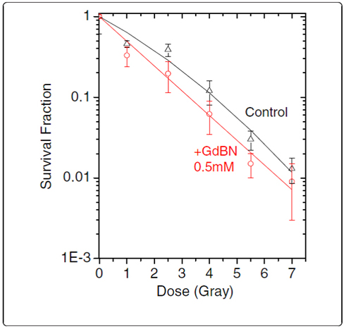
Finally, specific measurements with gamma rays have shown that the presence of Gadolinium-based nanoparticles in glioblastoma cells greatly increases their destruction. Researchers compared samples with or without GdBNs, and it turned out that there was a 23% improvement in the effect of radiation in the presence of NPs. This amplification appears to be linked to lysosome disturbance in cells that have incorporated NPs and not to cell respiration, since NPs do not enter mitochondria. Note that the SR-DUV microscopy studies have shown that the absorption of NPs is heterogeneous from one cell to another. This phenomenon is particularly interesting and a greater understanding of it would improve the protocol combining irradiation and the addition of NPs.
To conclude, the combination of microscopy techniques used by this group is a unique approach to observing the internalization of native (unlabeled) NPs in human cells and understanding better the role of nanoparticles as amplifiers of the effects of radiotherapy.