Bursting Bubbles – Tilted Holography of Magnetic Bubbles on SEXTANTS
Magnetic skyrmions are nanoscopic vortices of magnetisation, which carry the potential for exciting new applications in ultra-low energy advanced spintronic devices, due to their novel topological and transport properties.
An international team of researchers from SOLEIL (SEXTANTS beamline), The UK Skyrmion Project (Universities of Durham, Cambridge and Warwick, UK) and the University of Exeter, have used tilted resonant x-ray holography to investigate the exotic bound-pair skyrmion states known as biskyrmions, reported to exist in certain centrosymmetric ferromagnetic systems.
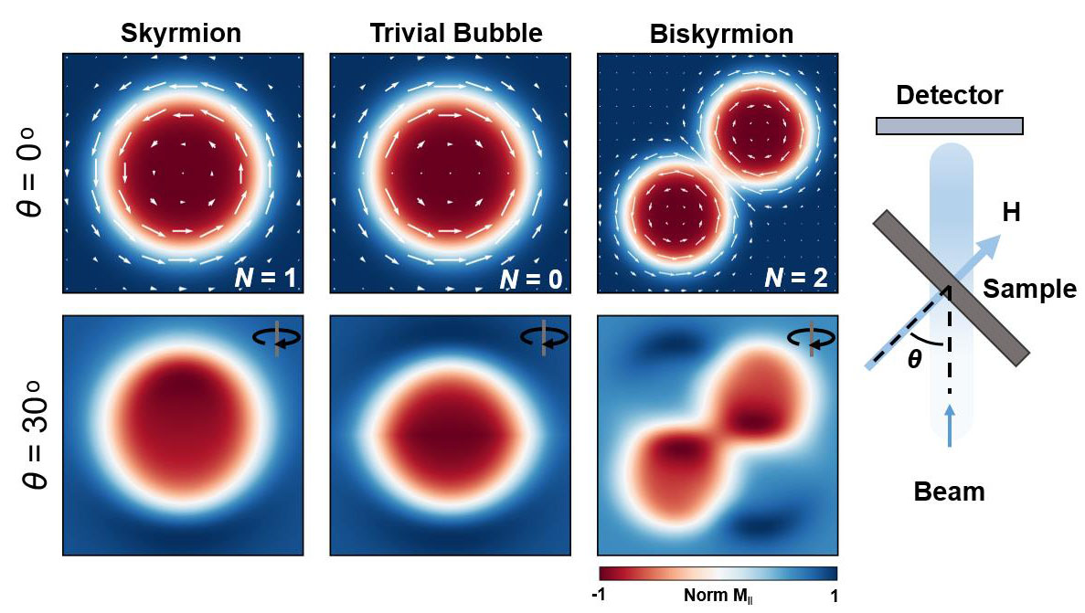
The results, published in ACS Nano, show that biskyrmions might not exist, suggesting previous reports have misidentified isolated magnetic bubble domains. By performing holographic imaging with tilted sample geometries, the study goes on to map the chirality of the domain walls forming these bubbles domains and determine that have a topologically trivial structure.
Initial reports of biskyrmions came from Lorentz transmission electron microscopy (LTEM) studies, a common electron-based imaging method, in which the deflection of electrons travelling through the magnetic field of a sample is used to image its magnetic structure. Whilst it boasts a high spatial resolution, the technique also maps the stray magnetic field of a sample, which has the potential to distort images and make interpretation more complex. In order to unambiguously determine the structure of these magnetisation textures, x-ray imaging was employed, which solely probes the magnetisation of a sample (Figure 1).
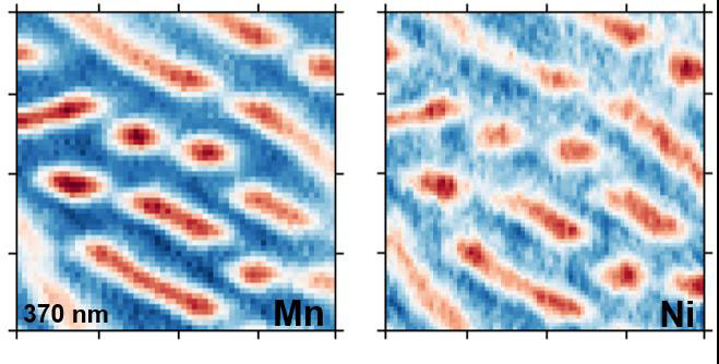
The researchers used the Fourier transform holography station at the SEXTANTS beamline to produce high-resolution x-ray images of the magnetic textures contained within the centrosymmetric ferromagnetic alloy MnNiGa. Magnetic contrast in such images is achieved by tuning the energy and polarization of the x-ray beam to resonance with electronic transitions in the magnetic ions of the sample, exploiting a phenomenon known as x-ray magnetic circular dichroism (XMCD). This also allows element specific imaging to be performed, and it was shown that the magnetic ordering on of the Mn and Ni sublattices is mutually ferromagnetic (Figure 2). As these element specific resonances occur at different energies, it is also possible to observe a shift in the spatial resolution of the images, corresponding to a difference in x-ray wavelengths. A spatial resolution of 23 ± 4 nm was attained at the Mn-L3 edge and 17 ± 4 nm at the Ni-L3 edge.
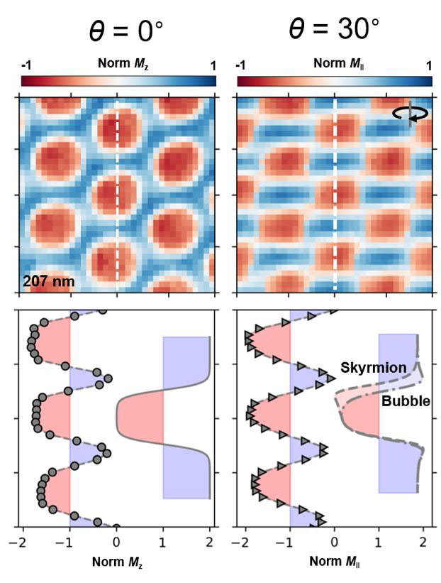
The single core bubbles observed were not consistent with the proposed biskyrmion structure; however, with images only projected normal to the sample plane, it was not possible to determine the winding direction of the bubble domain walls – a key characteristic in determining the topology in vortex-like states. By tilting the sample, a symmetric contraction of the bubbles consistent with topologically trivial bubbles was observed (Figure 3).
In a broader context, this work shows compelling evidence for the utility of resonant x-ray imaging methods in the field of nanoscale magnetism. The tilted projections point the way towards more advanced tomographic and laminographic imaging methods, which are the next step for magnetic imaging at synchrotrons.