Bacterial architects build network-like membrane structures to feed more efficiently
Press release - The cereal weevil, one of the world’s main crop pests, harbors symbiotic bacteria that live inside its cells. Scientists from INRAE and INSA Lyon, in collaboration with experts from the SOLEIL Synchrotron and Claude Bernard University in France, as well as the Max Planck Institute and EMBL in Germany, have discovered that these bacteria build complex, network-shaped membrane structures. These structures increase their surface area for exchange with the host cell, allowing the bacteria to absorb an essential nutrient: sugar. This is the first time that bacterial structures of this scale have been observed.
The results, published in Cell, open new research avenues for understanding microorganisms—particularly those living inside host cells—and offer promising new directions for combating crop-damaging insects.
The cereal weevil, one of the world’s main crop pests, harbors symbiotic bacteria that live inside its cells. Scientists from INRAE and INSA Lyon, in collaboration with experts from the SOLEIL Synchrotron and Claude Bernard University in France, as well as the Max Planck Institute and EMBL in Germany, have discovered that these bacteria build complex, network-shaped membrane structures. These structures increase their surface area for exchange with the host cell, allowing the bacteria to absorb an essential nutrient: sugar. This is the first time that bacterial structures of this scale have been observed. The results, published in Cell, open new research avenues for understanding microorganisms—particularly those living inside host cells—and offer promising new directions for combating crop-damaging insects.
The cereal weevil is one of the major pests affecting cereals such as wheat, rice, and maize, both in the field and in storage. It feeds directly on the grains, but it is not alone: it hosts symbiotic bacteria that live inside its cells. These bacteria, named Sodalis pierantonius, reside in large numbers within specialized insect cells. They provide the weevil with essential nutrients that are absent from its cereal-based diet. This is a mutually beneficial relationship: the bacteria use the sugars produced during the digestion of grains and, in return, supply the insect with essential nutrients such as vitamins and certain amino acids.
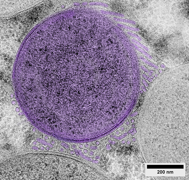
While scientists have long understood the importance of this exchange, its exact mechanisms remained unknown. To investigate, the researchers used electron microscopy with an advanced sample preparation method that preserves membranes more effectively. For the first time, the team observed original tubular patterns forming complex membrane structures built by the bacteria. To study the architecture and composition of these structures, the scientists developed new 3D microscopy and analytical methods using the STXM (Scanning Transmission X-ray Microscopy) technology, at the HERMES beamline of Synchrotron SOLEIL, in order to determine the carbon composition of samples with very high spatial resolution (30 nm).
Bacteria build an exchange network called the “tubenet”
The analyses revealed that the structures form a complex network of tubes, approximately 0.02 µm in diameter and several micrometers long, interconnecting multiple bacteria. Much like the microvilli1 of the human intestine, which increase the surface area available for nutrient absorption during digestion, these tubular structures enable bacteria to enlarge their exchange surface with the host cell and take up more sugar. In return, the bacteria produce essential nutrients for the host cell. The research team named these structures “Tubenets”—a contraction of tube and network—in reference to their shape.
While such surface-expanding structures are well known in multicellular organisms (such as the intestines or plant roots), this is the first time that a comparable structure has been identified in bacteria. It is possible that similar architectures exist in other bacterial species as well.
[1] Microvilli: Tiny, finger-like projections covering the cells of certain tissues such as those in the gallbladder or small intestine. They facilitate the absorption of external substances like nutrients.