A 300 keV cryo-electron microscope available at SOLEIL by 2025
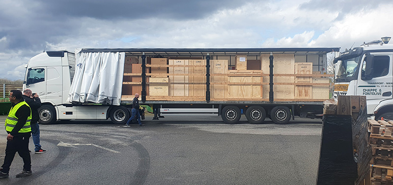
On Monday 18 March 2024, over 8 tons of equipment have been delivered to SOLEIL. These are parts for the new cryogenic electron microscope, which will be made available to users, on the basis of a call for projects, from 2025 onwards.
Cryo-electron microscopy (cryo-EM) is increasingly becoming a mainstream technology for studying the architecture of cells, viruses and protein assemblies from molecular to atomic resolution. Recent developments in microscope design and imaging hardware, paired with enhanced image processing and automation capabilities, appear poised to further advance the effectiveness of cryo-EM methods. These developments promise to increase the speed and extent of automation and to improve the resolutions that can be achieved, rendering this technology capable of determining a wide variety of biological structures.
The PIA3 - EQUIPEX+ FRANCE-CRYO-EM, a project to provide and operate top-end cryo-electron microscopy facilities for structural biology in France, was approved in December 2020. Finance was made available to equip Strasbourg, Grenoble and SOLEIL with 300 kV cryo-electron microscopes to operate ‘like synchrotron radiation beamlines’ (open, peer reviewed access). Further finance from regional CPER 2021-2027 funds was made available to equip a number of regional centers, including Paris-Saclay with modern 200 kV cryo-electron microscopes in order to suitably prepare projects to use time on the 300 kV instruments in the most efficient way. Hence the 200 kV microscope, currently being installed at the I2BC in Gif sur Yvette, and the 300 kV microscope at SOLEIL, will work in close synergy (Cryo-EM@Paris-Saclay consortium). The EQUIPEX+ project included 2 engineers who will share their time between the two instruments. The first of these, Eric Larquet, has been working 80% of his time at SOLEIL since April 2023. Recruitment of a second engineer is ongoing.
Access to the cryoEM at SOLEIL
Access for the structural biology community will initially be available via SUNSet, the SOLEIL’s platform to write and submit a proposal (https://www.synchrotron-soleil.fr/en/users), using a modified workflow in order to cater for the differences between an electron microscope (which can operate during storage ring shutdowns) and the synchrotron. It is expected that access will also be provided via the FRISBI portal (French Infrastructure for Structural Biology) via a mechanism currently being defined.
Expert users will come to carry out the first experiments in autumn 2024, followed by users who have responded to a call for projects from 2025 onwards.
Such an instrument represents a completely new technology at SOLEIL and is complementary to the current structural biology techniques and multimodal imaging methods already used at the synchrotron. This will allow SOLEIL users to cross the spatial scales of their biological samples from molecules to cell and tissues.
Setting up the microscope and its environment
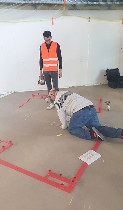
Consequently, a multi-disciplinary team has been assembled to define, design and construct a clean, low vibration, phonically insulated and electromagnetically shielded room in the space previously allocated for the beamline PROXIMA 2B (a second branch of the PROXIMA-2 beamline was to be built there, but it wasn’t eventually). Design work was completed in January 2024, and construction of the microscope room is advancing rapidly during storage ring shutdown periods. The room, to be built by CLIMASCIENCE, is expected to be completed and delivered in July 2024. The cryo-electron microscope, a ThermoFisher Scientific Titan KRIOS G4, will be pre-installed in May and June 2024, and ‘switched on’ following the completion of the microscope room.
Acceptance tests of the KRIOS microscope took place in January, at the ThermoFisher Scientific site. Eric Larquet and Pierre Legrand, scientist in charge of PROXIMA-1 beamline and of the cryoEM project, witnessed the test measurements which demonstrated an excellent performance, including the determination of the structure of the protein apo-ferritin* to almost 1.11 Angstrom resolution – a world record resolution at that time.
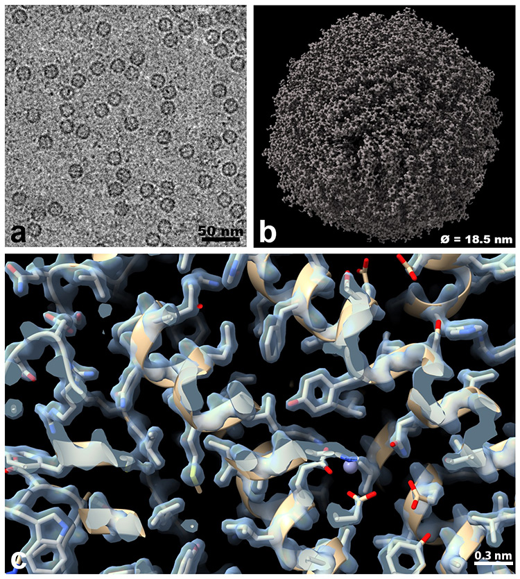
a. Representative electron micrograph of apoferritin* molecule in vitreous ice – each small circular shape corresponds to an apoferritin molecule..
b. 3D Cryo-EM density map of a single apoferritin molecule (diameter: 18.5 nm) reconstructed from 2D projections.
c. Fitting atomic models of amino acid chains in the Cryo-EM density map (Resolution: 1.26 Å).
On Monday 18th March at 04h30, the lorry carrying SOLEIL’s microscope left the Thermo Fisher Scientific Eindhoven factory in order to arrive at SOLEIL at midday in order to cause minimum disruption to the program of machine physics work on the storage ring. Twenty large crates, a weight in excess of 8 tons of equipment, had to be transported, via Fenwick, from the lorry to a storage area in front of the ODE beamline in under 2 hours. Thanks to the intervention of Cyrille Azambourg, Victor Filipe (SOLEIL’s Bât-Infra group) and staff from DEMECO, the lorry was unloaded rapidly and efficiently.
The delivery marks the start of a new era for structural biology at SOLEIL, and its commissioning is eagerly awaited by our user community.
* Apoferritin is a 24-subunit protein (474 kDa), considered as the standard for cryo-EM single particle analysis, used for quality control; this protein is the cryoEM equivalent of the lysozyme used in protein crystallography.