PUMA
PUMA (French for “Photons Utilisés pour les Materiaux Anciens”) is a hard X-ray beamline optimized for the scientific communities of the heritage sciences but not exclusive to them. Our microbeam endstation features a 5 (v) x 7 (h) µm2 spot size, in an energy range from 4 to 22 keV. Available experimental techniques include X-ray fluorescence spectroscopy (XRF), X-ray absorption spectroscopy (XANES), X-ray diffraction (XRD) and X-ray excited optical luminescence spectroscopy (XEOL).
The PUMA beamline opened its doors to users in February of 2019. The beamline has been built and optimized for the heritage science community and most user experiments submitted are from the fields of Archaeology, Paleontology, Paleo-Environments, Art History and Conservation Sciences. However, the beamline is open to all users and has hosted successfully experiments from Environmental Sciences, Earth Sciences, Biology, Soft-Matter and Medicine.
Our working horse is a microbeam endstation, which produces a 5 µm (vertical) x 10 µm (horizontal) spot size. The sample is typically mounted in a 45 degree geometry towards the incoming beam, with the SDD detector for the XRF signal at 90 degrees to the incoming beam. A video-microscope allows to image the sample surface at all time in the geometry. The image is calibrated on the beam position and allows to align the surface features of the sample with a precision of a few micrometers with respect to the beam.
A Si(111) double crystal monochromator with fixed-exit geometry allows to select the energy in a range from 4 to 22 keV. A diamond intensity monitor at the exit of the KB mirror system allows for constant monitoring of the beam intensity.
XRF and XANES can be performed using the silicon drift detec
tor. Currently it is not possible to perform XAS in transmission mode.
An hybrid pixel detector (ImXpad) can be mounted behind the sample to collect X-ray diffraction patterns in transmission mode only.
The microscope can be used to collect the XEOL signal from the sample, either in imaging mode, using the camera of the microscope or in spectroscopy mode, using a full spectrometer.
This endstation supports to sample stages. A set of high precision stages with submicroscopic resolution for centimetric samples and a set of larger stages for heavier samples tens of centimeters in length. All stages support continuous scanning modes for fast and distortion-free acquisition.
The setup is in air, which gives us a lot of flexibility to mount different setups. If you want to bring your own equipment to the beamline, please contact the beamline staff.
A 3D microtomography endstation is currently in development, but not yet available for user.

The beamline has been constructed and will be operated in collaboration with the IPANEMA laboratory. In the future, it will contribute researchers and engineers to the PUMA staff.
Acknowledgments
The construction of the PUMA beamline has been financed by the region île-de-France and the French state through the CPER 20017 - 2013 program.
The equipment of the micro focusing station is property of the CNRS and has been financed by the PATRIMEX equipped grant in the framework of the “Investissements d'Avenir” program of the “Agence Nationale de la Recherche” (ANR).

Beamline news
All PUMA's News
SOLEIL Highlights 2025
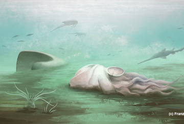
The "oldest octopus"'s mistaken identity unmasked using the PUMA beamline
Team

Technical data
- Energy Domain
-
4 - 22 keV micro beam endstation
4 - 60 keV full field endstation
- Source
-
Wiggler W164.2mm×20 periods, B = 1.8 T, medium section
- Beam modes / monochromators
-
Monochromatic beam : Double crystal monochromator (DCM) Si111 (4-20 keV) and Si220 (20-60 keV)
- Optics
-
Diaphragm 1.45×2.8 mm² (H×V) @ 17 m
Beryllium window : Be 300 µm @ 19 m
Removable filter : CVD 1000 µm @ 19 m
Primary slits @ 19.5 m
Horizontal mirror @ 21 m : θ = 1.3 mrad fixe, longeur focale variable.
DCM @ 22.5 m : Si 111 ou Si 220
Secondary slits @ 24 m
Tertiary slits @ 40 m
KB mirror @ 63 m : B4C 60 nm ou Rh 60 nm layer
Sample @ 63.3 m
- Experimental techniques
-
X-ray fluorescence spectroscopy (XRF)
X-ray absorption spectroscopy (XANES)
X-ray powder diffraction (XRD) - transmission geometry only
X-ray excited optical luminescence specroscopy (XEOL)
- Beam size on the sample Micro beam with KB mirror system : 5 µm x 7 µm (VxH)
-
Micro beam with KB mirror system : 5 µm x 7 µm (VxH)
Full field : Max. field of view 20 mm x 12 mm (HxV)
Scientific opportunities
- Archeology
- Paleontology
- Paleo-environments
- Conservation sciences
- Art History
- Radiation effects
- Biology
- Soft Matter
- Environmental Sciences
- Geosciences