Simultaneous
fast-scanning XRF,
dark field, phase-,
and absorption contrast
tomography
The multi-technique “FLYSCAN”
data acquisition scheme developed
at Synchrotron SOLEIL permits to perform
fast continuous scanning making scanning
tomography feasible during typical user
experiments. Here we present the recent
results of such simultaneous hard X-ray
multi-technique tomography. This fast
scanning scheme will be implemented
at the Nanoscopium beamline for large field
of view 2D and 3D multimodal imaging.
➊
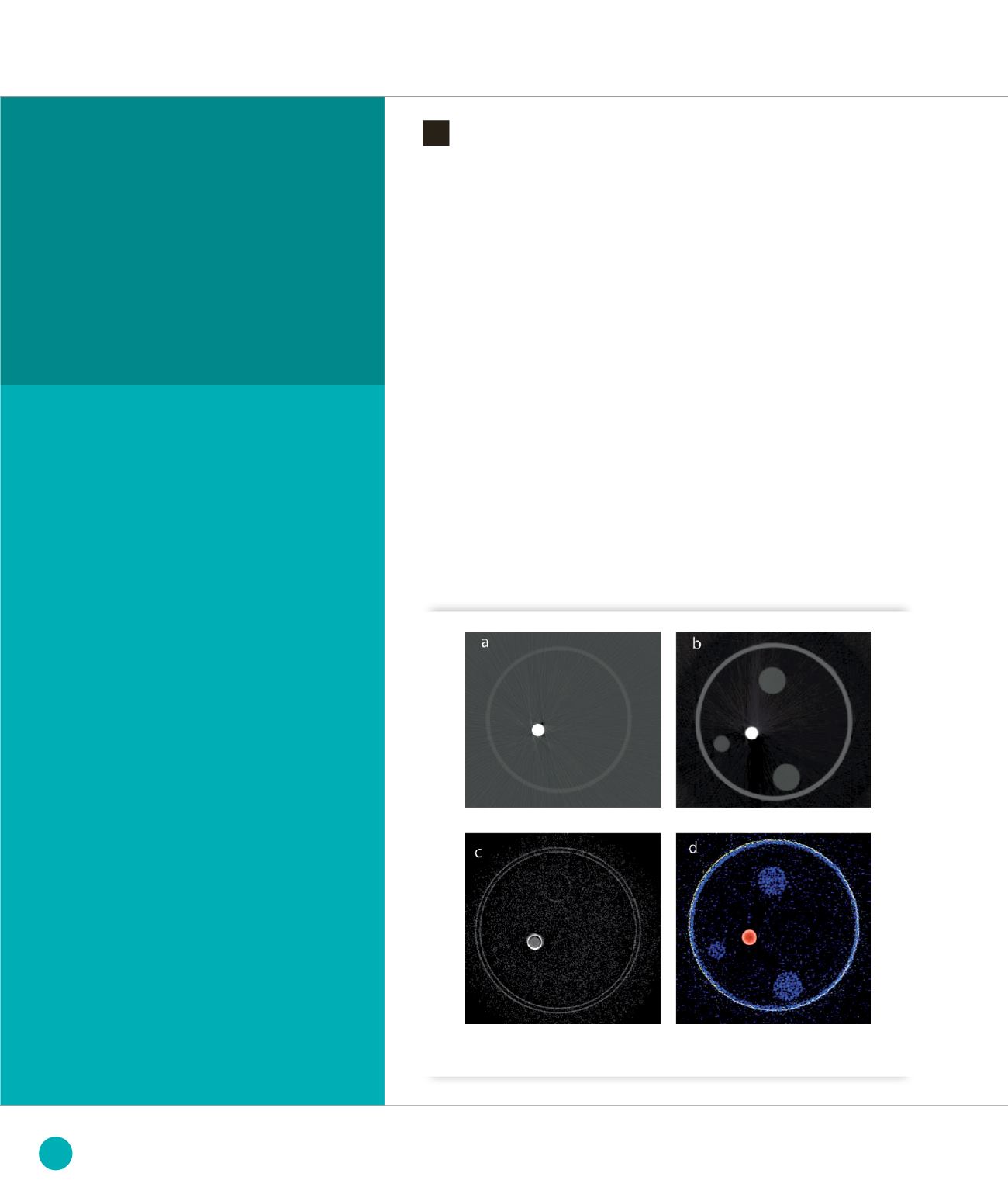
Multimodal tomograms :
a
) Attenuation,
b
) Phase,
c
) Dark field, d) X-Ray Fluorescence (red: copper, yellow: silicon,
blue : compton scattering)
Multi-technique scanning hard X-ray
imaging is a powerful technique to obtain
spatially resolved and high sensitivity
quantitative elemental and structural
information by raster scanning the sample
in the focused X-ray beam while measuring
the transmitted beam and/or the secondary
radiation emitted by the sample.
Complementary contrast mechanisms
such as absorption, phase, dark field and
X-ray fluorescence can be measured
simultaneously for obtaining information
on the sample structure, composition
and chemistry. The combination of all
these contrast modalities by scanning
tomography provides a unique tool for
the nondestructive quantitative study
of the sample in three dimensions.
The Nanoscopium beamline, (under
construction), is dedicated to scanning
hard X-Ray 2D/3D multimodal imaging
in the 5 to 20 keV energy range. The
beamline aims to provide a stable and high
intensity nano-beam which can be tailored
in the 30-500 nm size-range depending
on the experimental needs. The study of
heterogeneous, fragile or large samples
will be assured by fast and continuous
scanning with sensitive and high frame
rate detectors.
The multi-technique “FLYSCAN” data
acquisition scheme [1] developed at
SOLEIL permits to perform fast continuous
multi-technique scanning. In this
paper, we present the proof of principle
experiments of scanning tomography
by the FLYSCAN scheme together with
volumetric reconstruction, performed by
dedicated algorithm to each measured
contrast modality.
The experiments were carried out at the
Metrologie beamline of SOLEIL using a
temporary scanning microprobe set-up.
Monochromatic X-rays of 14 keV were
focused down to 1
µ
m by a Fresnel Zone
Plate. The read-out of a silicon photodiode
(incoming beam intensity), pixel array
detector (XPAD) (transmitted beam), a
silicon drift detector (X-ray fluorescence)
and the motor encoders were hardware
synchronized by a TTL signal (Master
Trigger).
MODELING, METHODOLOGY AND INSTRUMENTATION
126
SOLEIL
HIGHLIGHTS
2013


