NANOSCOPIUM beamline
& Informatics-Electronics Group
ASSOCIATED PUBLICATION
Simultaneous fast scanning XRF, dark field,
phase-, and absorption contrast tomography
K. Medjoubi*, A. Bonissent, N. Leclercq,
F. Langlois, P. Mercère and A. Somogyi
Proceeding: X-Ray Nanoimaging: Instruments
and Methods, San Diego (US), August 25, 2013,
Proceedings of SPIE, 2013, 8851:art.n° 88510P
* Synchrotron SOLEIL, l’Orme des Merisiers,
St Aubin BP48, 91192 Gif sur Yvette Cedex,
France
CORRESPONDING AUTHOR
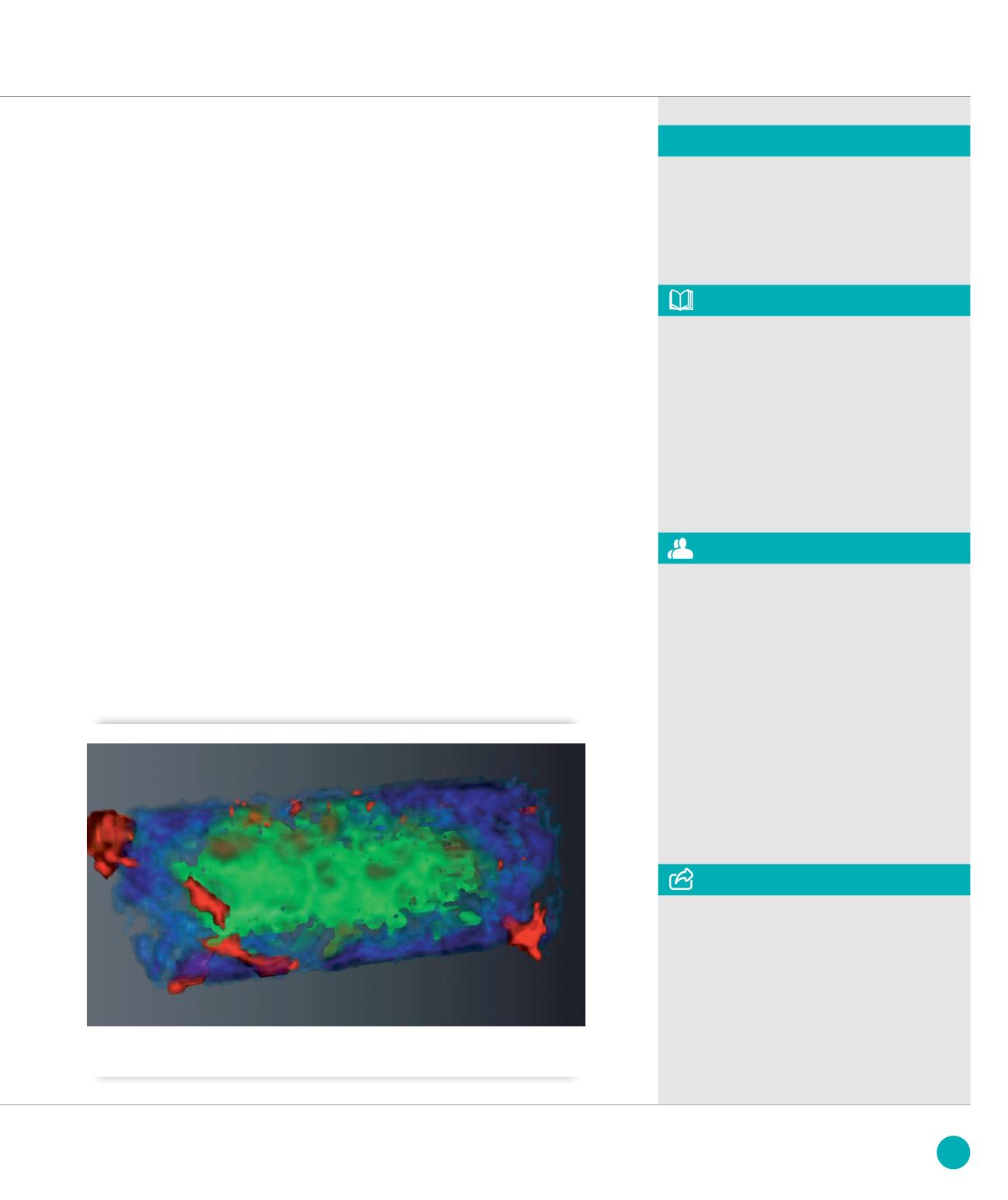
➋
Volume rendering of the 3D tomograms combining the sample absorption (green), the Ca (blue) and the Fe (red)
distributions.
REFERENCE
[1] Medjoubi et al. J. Synchrotron Rad.
20 (2013), 293
The tomography of a slice of a test sample
(glass capillary containing two nylon fibers
and a copper wire) was performed with
500 angular projections, over the full 360°
range, in less than 1 hour total acquisition
time which was limited by the flux
available on the bending magnet beamline.
Figure
➊
presents the reconstructed
tomogram of each contrast modality. The
conventional transmission CT (fig.
➊
.
a
)
reveals the copper wire and with lower
contrasts the capillary itself. The phase
tomogram (fig.
➊
.
b
), where the nylon
fibers are visible with a very good contrast,
provides complementary information about
light materials. The dark field contrast
(fig
➊
.
c
) shows the capillary walls and
the wires. In the XRF tomogram shown
in fig
➊
.
d
the reconstructed Si (capillary),
Cu and Compton signals are overlapped.
3D imaging was performed on an
80 µm-thick polished geological sample
section consisting of calcium carbonate
and organic material layers. The sample
also contained small pyrite crystals. The
2D projection images, of 300 x 10 pixels
(pixel size: 3 x 3 µm
2
), were recorded at
6° rotational intervals over 360°. The total
acquisition time was less than 2 hours.
Figure
➋
presents the volume rendering of
the absorption, Fe and Ca reconstructions.
The pyrite clusters having high Fe content,
can be clearly identified.
The performance and the potential
of the FLYSCAN architecture for fast multi-
technique scanning tomographic imaging
have been demonstrated. Simultaneous
acquisition of 3D tomographic data sets
of X-ray fluorescence, transmission,
phase contrast and dark field has been
performed in 1-2 hours total acquisition
time using a prototype FZP-based
microprobe set-up. This experiment
performed in step scan mode in the
same experimental conditions would
take several days.
At the Nanoscopium beamline, where
the available flux will be 10
3
-10
4
times
higher than at the Metrology beamline,
the presented experiments will be done
in less than half an hour. This will
permit the use of scanning tomography
techniques routinely during a user
experiment.
The authors are grateful to Pascal Phillipot
and Marie Sforna from the Institut de
Physique du Globe de Paris for providing
and preparing the geological sample. We
acknowledge G. Baranton (Nanoscopium),
P. Da Silva (Metrology) and our colleagues
from the Informatics and Electronics
(ICA/ECA) Support Groups of SOLEIL
who helped with the test experiments.
127
SOLEIL
HIGHLIGHTS
2013


